Review
. 2006 May 31;6(1):33-42.
doi: 10.1102/1470-7330.2006.0007.

New MR techniques for the detection of liver metastases

Affiliations
Review

New MR techniques for the detection of liver metastases

J Ward. Cancer Imaging. .

Abstract

It is well established that hepatic resection improves the long-term prognosis of many patients with liver metastases. However, incomplete resection does not prolong survival, so knowledge of the exact extent of intra-hepatic disease is crucially important in determining patient management and outcome. MR imaging is well recognised as one of the most sensitive methods for detecting metastases. Recent developments in gradient coil design, the use of body phased array coils and the availability of novel MR contrast agents have resulted in MR being recognised as the pre-operative standard in this group of patients. However, diagnostic efficacy is extremely dependent on the choice and optimisation of pulse sequences and the appropriate use of MR contrast agents. This article reviews current MR imaging techniques for the detection and characterisation of metastases and discusses the relative capability of different techniques for detecting small lesions.


Figures

Figure 1
Figure 1
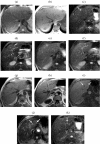
Relative liver-to-lesion contrast on unenhanced breathhold (BH) imaging. BH T2w FSE (a) illustrating poor visibility of multiple metastases. The lesions are more conspicuous on the corresponding IPT1w images. (b) In a second patient also with multiple small metastases, two left lobe lesions (arrows) are highly conspicuous on BH STIR ((c) and (d)). Both lesions are visible on BH FSE ((e) and (f)) and IPT1w ((g) and (h)) but with reduced liver-to-lesion contrast compared with STIR. Two additional sub-cm lesions (arrows) are well seen on SPIO-enhanced 3D FS T1w (i) and T2w GRE images (j) but only one is seen on the corresponding STIR image (k).
Figure 2
Figure 2
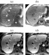
Dynamic Gd-enhanced MR versus unenhanced MR for lesion detection—role of 3D FS T1w GRE imaging. In a patient with pancreatic cancer several metastases larger than 1 cm are well seen on BH T2 FSE (a) and IPT1w (b) images. Adjacent thin-slice (2.5 mm) portal phase post-Gd 3D FS T1w GRE images ((c) and (d)) obtained at the same level as ((a) and (b)) clearly show several additional previously undetected sub-cm metastases.
Figure 3
Figure 3
Value of arterial phase for characterising and delineating hypovascular metastases. Characteristic continuous rim enhancement is clearly seen on arterial phase post-Gd 3D FS T1w GRE images (a). The lesions are still conspicuous by the portal phase (b) but the enhancing rim is less apparent and the lesions appear smaller.
Figure 4
Figure 4
Role of Gd-enhanced delayed imaging with fat suppression for detecting small metastases on the liver surface. In a patient with colorectal cancer small surface deposits (arrows) are well seen on 3D FS T1w GRE images obtained approximately 10 min after Gd ((a) and (b)). The lesions are not visible on the earlier portal phase images ((c) and (d)). Surface lesions are also highly conspicuous on FS T2w GRE images following SPIO ((e) and (f)).
Figure 5
Figure 5
Multiple metastases—improved detection with Gd-EOB-DTPA at the hepatocyte phase of enhancement. Compared with non-contrast T1w images (a) liver-to-lesion contrast is improved on 20 min post-contrast T1w images (b). Additional surgically confirmed sub-cm lesions (arrows) were only visible on hepatocyte phase images ((c) and (d)). Adapted with permission from Robinson PJA, Ward J (2006) MRI of the Liver: A Practical Guide. Informa Healthcare, New York.
Figure 6
Figure 6
Improved detection of metastases with 24 h mangafodipir-enhanced imaging. Multiple metastases (arrows) are seen with high lesion-to-liver contrast on 20 min post-mangafodipir T1w 2D GRE images (a). However several additional lesions are only visible on the corresponding images obtained 24 h after contrast (b) when the background liver signal has returned to normal, due to retained contrast in the compressed liver tissue at the periphery of the lesions. (Images courtesy of Dr J. Healy.)
Figure 7
Figure 7
Improved detection of small metastases with optimised SPIO-enhanced T2w GRE imaging. In a patient with colorectal metastases and a fatty liver, right and left lobe lesions (arrows) are well seen on HASTE (a) and IPT1w (b) images. The lesions are isointense against the reduced signal of the adjacent fatty liver on OPT1w (c). Additional small metastases (arrows) not seen on (a–c) are clearly seen on SPIO-enhanced T2w GRE images (d) acquired with a 6 mm slice thickness and fat suppression.
Figure 8
Figure 8
Value of dynamic high-resolution T1w imaging with ferucarbotran for depicting small metastases. Multiple small metastases (many not visible on unenhanced images) are highly conspicuous on 3D FS T1w GRE imaging (effective slice thickness 2.5 mm) obtained 45 s after bolus injection of ferucarbotran ((a) and (b)). Note the relatively weak T1 effect resulting in isointensity of the background liver and vessels. The lesions are also well seen on corresponding T2w GRE images (c–e) obtained 10 minutes after (a) and (b).

References

    1. Malafosse R, Penna C, Sa Cunha A, Nordlinger B. Surgical management of hepatic metastases from colorectal malignancies. Ann Oncol. 2001;12:887–94. - PubMed
    1. Miyazaki M, Ito H, Nakagawa K, Ambiru S, Shimizu H, Okuno A, et al. Aggressive surgical resection for hepatic metastases involving the inferior vena cava. Am J Surg. 1999;177:294–8. - PubMed
    1. Lodge JP, Ammori BJ, Prasad KR, Bellamy MC. Ex vivo and in situ resection of inferior vena cava with hepatectomy for colorectal metastases. Ann Surg. 2000;231:471–9. - PMC - PubMed
    1. Jaeck D, Bachellier P, Guiguet M, Boudjema K, Vaillant JC, Balladur P, et al. Long-term survival following resection of colorectal hepatic metastases. Br J Surg. 1997;84:977–80. - PubMed
    1. Harrison LE, Brennan MF, Newman E, Fortner JG, Picardo A, Blumgart LH, et al. Hepatic resection for noncolorectal, nonneuroendocrine metastases: a fifteen-year experience with ninety-six patients. Surgery. 1997;121:625–32. - PubMed

MeSH terms

LinkOut - more resources