. 2006 Jun 27;103(26):9964-9.
doi: 10.1073/pnas.0603507103. Epub 2006 Jun 9.

Nucleophosmin/anaplastic lymphoma kinase (NPM/ALK) oncoprotein induces the T regulatory cell phenotype by activating STAT3

Affiliations

Nucleophosmin/anaplastic lymphoma kinase (NPM/ALK) oncoprotein induces the T regulatory cell phenotype by activating STAT3

Monika Kasprzycka et al. Proc Natl Acad Sci U S A. .

Abstract

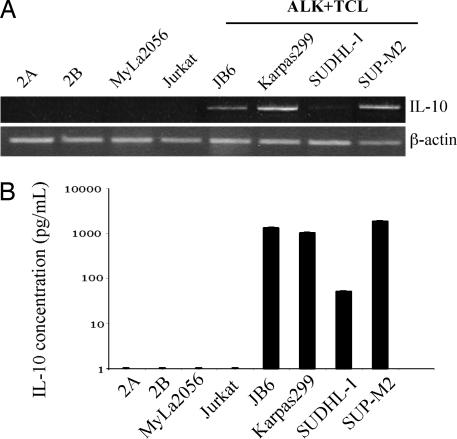
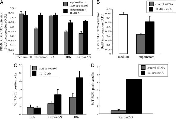
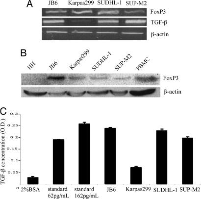
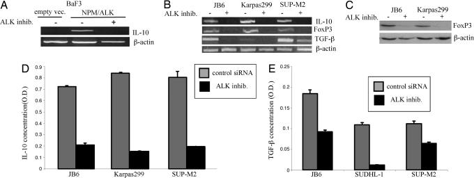
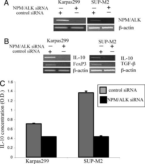
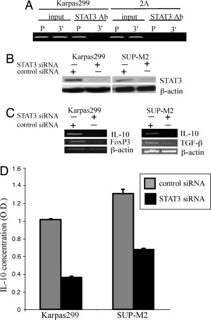
The mechanisms of malignant cell transformation mediated by the oncogenic, chimeric nucleophosmin/anaplastic lymphoma kinase (NPM/ALK) tyrosine kinase remain only partially understood. Here we report that the NPM/ALK-carrying T cell lymphoma (ALK+TCL) cells secrete IL-10 and TGF-beta and express FoxP3, indicating their T regulatory (Treg) cell phenotype. The secreted IL-10 suppresses proliferation of normal immune, CD3/CD28-stimulated peripheral blood mononuclear cells and enhances viability of the ALK+TCL cells. The Treg phenotype of the affected cells is strictly dependent on NPM/ALK expression and function as demonstrated by transfection of the kinase into BaF3 cells and inhibition of its enzymatic activity and expression in ALK+TCL cells. NPM/ALK, in turn, induces the phenotype through activation of its key signal transmitter, signal transducer and activator of transcription 3 (STAT3). These findings identify a mechanism of NPM/ALK-mediated oncogenesis based on induction of the Treg phenotype of the transformed CD4(+) T cells. These results also provide an additional rationale to therapeutically target the chimeric kinase and/or STAT3 in ALK+TCL.


Conflict of interest statement

Conflict of interest statement: No conflicts declared.

Figures

Fig. 1.
Fig. 1.
IL-10 expression by ALK+TCL cells. IL-10 expression in ALK+TCL cell lines (JB6, Karpas299, SUDHL-1, and SUP-M2), T cell lymphoma cell lines derived from lymphomas involving skin (2A, 2B, and MyLa2056), and T cell lymphoblastic lymphoma (Jurkat) detected at the mRNA level by RT-PCR (A) and the protein level by EIA (B).
Fig. 2.
Fig. 2.
ALK+TCL-derived IL-10 is immunosuppressive and enhances survival of the ALK+TCL cells. (A) Anti-IL-10 antibody inhibits immunosuppressive activity of the ALK+TCL cell supernatants. Inhibitory effect of the ALK+TCL cell line (JB6 and Karpas299) supernatants pretreated with the anti-IL-10 antibody or the isotype-matched antibody on proliferation of the T cell rich PBMCs stimulated by the anti-CD3/anti-CD28 antibody tandem. Medium alone, medium with added recombinant IL-10, and a supernatant from the IL-10 nonsecreting 2A ALK−TCL cell line served as additional controls. (B) siRNA-mediated depletion of IL-10 inhibits immunosuppressive activity of the ALK+TCL cell supernatants. Inhibitory effect of the supernatant from the IL-10 siRNA- vs. nonsense siRNA-treated Karpas299 cells on proliferation of the anti-CD3/anti-CD28 antibody-stimulated PBMCs. (C) Proapoptotic effect of the anti-IL-10 antibody on the ALK+TCL cells. Viability of the Karpas299 and JB6 cell lines cultured in the presence of the anti-IL-10 or isotype-matched antibody was determined by the DNA fragmentation (TUNEL) assay. ALK−TCL 2A cell line served as an additional control. (D) Proapoptotic effect of the IL-10 siRNA on the ALK+TCL cells. Viability of the Karpas299 cell line treated with IL-10-specific or nonsense siRNA as determined by the TUNEL assay.
Fig. 3.
Fig. 3.
ALK+TCL cells display Treg cell phenotype. Expression of mRNA coding for FoxP3 and TGF-β (A) and FoxP3 protein (B) and the secreted TGF-β protein (C) by the ALK+TCL lines.
Fig. 4.
Fig. 4.
Expression of IL-10, FoxP3, and TFG-β are induced by NPM/ALK expression and enzymatic activity. (A) Expression of IL-10 mRNA in BaF3 cells transfected with empty vector and NPM/ALK before and after treatment with an ALK inhibitor WHI-P154 (10). (B) Expression of IL-10, FoxP3, and TGF-β mRNA in the depicted ALK+TCL cell lines before and after treatment with the ALK inhibitor. (C) Expression of FoxP3 protein in the ALK+TCL cell lines before and after treatment with the ALK inhibitor. (D) Concentration of IL-10 in the ALK+TCL cell line supernatant before and after cell treatment with the ALK inhibitor. (E) Concentration of TGF-β in the ALK+TCL cell line supernatant before and after cell treatment with the ALK inhibitor.
Fig. 5.
Fig. 5.
siRNA-mediated NPM/ALK depletion results in loss of the Treg phenotype. Two ALK+TCL cell lines, Karpas299 and JB6, were treated with the NPM/ALK-specific or control, nonsense siRNA and evaluated for expression of NPM/ALK (A), IL-10, FoxP3, and TGF-β (B), and the secreted IL-10 protein (C).
Fig. 6.
Fig. 6.
NPM/ALK induces the Treg phenotype through STAT3. (A) Binding of STAT3 to the IL-10 gene promoter. Protein cell lysates from the IL-10-expressing, ALK+TCL cell line Karpas299 and ALK−TCL cell line 2A were analyzed in the chromatin immunoprecipitation (ChIP) assay by using an anti-STAT3 antibody and primer pairs specific for promoter (P) and 3′ end (3′) of the IL-10 gene. Nonimmunoprecipitated lysates (input) served as positive controls. (B) Effect of the STAT3 siRNA on STAT3 expression. Two ALK+TCL cell lines, Karpas299 and SUP-M2, were treated with STAT3-specific and control, nonspecific siRNA and evaluated for expression of the STAT3 protein. (C) Effect of the siRNA-mediated STAT3 depletion on the Treg phenotype. Karpas299 (Left) and SUP-M2 (Right) were treated with STAT3-specific and nonspecific siRNA and evaluated for expression of the IL-10, FoxP3, and TGF-β mRNA. (D) Effect of the siRNA-mediated STAT3 depletion on expression of the secreted IL-10. Supernatants form the Karpas299 and SUP-M2 pretreated with STAT3 and nonspecific siRNA were analyzed for IL-10 concentration in an EIA assay.

Comment in

  • STAT3: a multifaceted oncogene.
    Levy DE, Inghirami G. Levy DE, et al. Proc Natl Acad Sci U S A. 2006 Jul 5;103(27):10151-10152. doi: 10.1073/pnas.0604042103. Epub 2006 Jun 26. Proc Natl Acad Sci U S A. 2006. PMID: 16801534 Free PMC article. No abstract available.

References

    1. Wasik M. A. Am. J. Clin. Pathol. 2002;118:S81–S92. - PubMed
    1. Shiota M., Fujimoto J., Semba T., Satoh H., Yamamoto T., Mori S. Oncogene. 1994;9:1567–1574. - PubMed
    1. Morris S. W., Naeve C., Mathew P., James P. L., Kirstein M. N., Cui X., Witte D. P. Oncogene. 1997;14:2175–2188. - PubMed
    1. Fujimoto J., Shiota M., Iwahara T., Seki N., Satoh H., Mori S., Yamamoto T. Proc. Natl. Acad. Sci. USA. 1996;93:4181–4186. - PMC - PubMed
    1. Bischof D., Pulford K., Mason D. Y., Morris S. W. Mol. Cell. Biol. 1997;17:2312–2325. - PMC - PubMed

Publication types

MeSH terms