. 2006 Jun 20;103(25):9703-8.
doi: 10.1073/pnas.0601940103. Epub 2006 Jun 9.

Circadian organization of the mammalian retina

Affiliations

Circadian organization of the mammalian retina

Guo-Xiang Ruan et al. Proc Natl Acad Sci U S A. .

Abstract

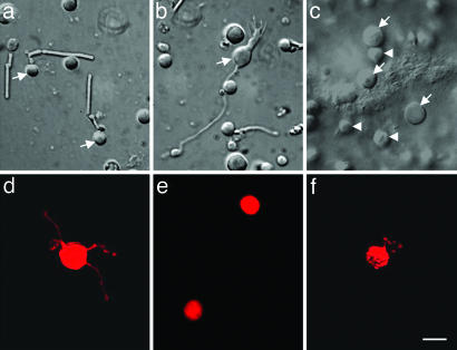
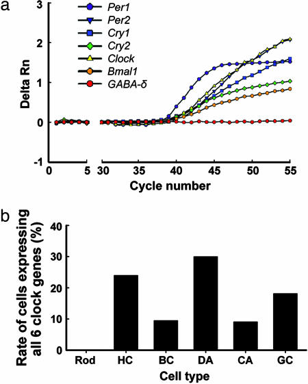
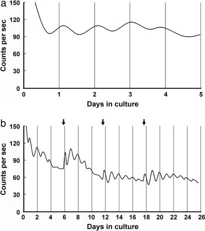
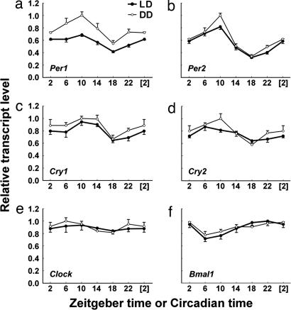
The mammalian retina contains an endogenous circadian pacemaker that broadly regulates retinal physiology and function, yet the cellular origin and organization of the mammalian retinal circadian clock remains unclear. Circadian clock neurons generate daily rhythms via cell-autonomous autoregulatory clock gene networks, and, thus, to localize circadian clock neurons within the mammalian retina, we have studied the cell type-specific expression of six core circadian clock genes in individual, identified mouse retinal neurons, as well as characterized the clock gene expression rhythms in photoreceptor degenerate rd mouse retinas. Individual photoreceptors, horizontal, bipolar, dopaminergic (DA) amacrines, catecholaminergic (CA) amacrines, and ganglion neurons were identified either by morphology or by a tyrosine hydroxylase (TH) promoter-driven red fluorescent protein (RFP) fluorescent reporter. Cells were collected, and their transcriptomes were subjected to multiplex single-cell RT-PCR for the core clock genes Period (Per) 1 and 2, Cryptochrome (Cry) 1 and 2, Clock, and Bmal1. Individual horizontal, bipolar, DA, CA, and ganglion neurons, but not photoreceptors, were found to coordinately express all six core clock genes, with the lowest proportion of putative clock cells in photoreceptors (0%) and the highest proportion in DA neurons (30%). In addition, clock gene rhythms were found to persist for >25 days in isolated, cultured rd mouse retinas in which photoreceptors had degenerated. Our results indicate that multiple types of retinal neurons are potential circadian clock neurons that express key elements of the circadian autoregulatory gene network and that the inner nuclear and ganglion cell layers of the mammalian retina contain functionally autonomous circadian clocks.


Conflict of interest statement

Conflict of interest statement: No conflicts declared.

Figures

Fig. 1.
Fig. 1.
Six types of mouse retinal cells were identified and harvested for RT-PCR. Rods (a; arrows) and rod bipolar cells (b; arrow) were identified in dispersed cultures based on morphology. Horizontal cells (c; arrows) were identified and harvested from whole-mount rd mouse retinas. The relatively small surface cells (arrow heads) are either bipolar cells or remaining photoreceptors. DA cells (d) and type 2 CA cells (e) were identified in dispersed cultures of TH::RFP transgenic mouse retinas by RFP fluorescence. Ganglion cells (f) were identified in dispersed cultures by DiI fluorescence after injection and retrograde transport from the lateral geniculate nuclei. (Scale bar: 10 μm.)
Fig. 2.
Fig. 2.
Simultaneous detection of all six core clock genes in individual retinal neurons. (a) Real-time PCR amplification plot of a representative DA cell in which all six clock gene transcripts, but not the GABA-δ receptor subunit transcript (a negative control gene in DA cells), were detected. Data are presented as the average of duplicate amplifications for each gene. Detection of specific PCR products was monitored as the relative fluorescence (delta Rn) in the intensity of reporter dye. (b) The percentage of cells expressing all six core clock genes in each cell type is shown. Rod, rods; HC, horizontal cells; BC, rod bipolar cells; DA, dopaminergic amacrine cells; CA, type 2 catecholamine amacrine cells; GC, ganglion cells.
Fig. 3.
Fig. 3.
Real-time monitoring of PER2::LUC expression in isolated rd mouse retinas. (a) Representative record showing retinal rhythms during the initial 5 days of culture. Retinas were dissected and explanted just before lights off (ZT 12). Vertical lines indicate the projected time of lights off on each day in DD (projected ZT 12). Photon counts per second are plotted against days in culture. (b) Long-term culture of an rd mouse retina showing persistent circadian rhythms in PER2::LUC expression. Arrows indicate times of medium changes.
Fig. 4.
Fig. 4.
Temporal expression profiles of Per1 (a), Per2 (b), Cry1 (c), Cry2 (d), Clock (e), and Bmal1 (f) in rd mouse retinas under LD (●) and DD (○) conditions. Data are presented as means ± SEM from five mice and expressed as a percentage of the maximum mean expression. Points at ZT/CT [2] are duplicates of ZT/CT 2 replotted to show 24-h trends. One-way ANOVA revealed P < 0.001 for Per1 and Per2 under both LD and DD conditions, P < 0.01 for Cry1 and Bmal1 under both LD and DD conditions, P < 0.05 for Cry2/LD, and P < 0.001 for Cry2/DD. For Per1, Per2, Cry1, Cry2, and Bmal1 expression, the peak-trough amplitudes under LD conditions were 1.65-, 2.54-, 1.46-, 1.35-, and 1.39-fold, respectively, whereas, under DD conditions, they were 1.84-, 2.95-, 1.50-, 1.73-, and 1.26-fold, respectively.

References

    1. LaVail M. M. Science. 1976;194:1071–1074. - PubMed
    1. Terman J. S., Reme C. E., Terman M. Brain Res. 1993;605:256–264. - PubMed
    1. Grace M. S., Wang L. M., Pickard G. E., Besharse J. C., Menaker M. Brain Res. 1996;735:93–100. - PubMed
    1. Tosini G., Menaker M. Science. 1996;272:419–421. - PubMed
    1. Tosini G., Menaker M. Brain Res. 1998;789:221–228. - PubMed

Publication types

LinkOut - more resources