. 2006 Jul;141(3):1068-77.
doi: 10.1104/pp.106.081463. Epub 2006 Jun 9.

Biosynthesis of ascorbic acid in legume root nodules

Affiliations

Biosynthesis of ascorbic acid in legume root nodules

Manuel A Matamoros et al. Plant Physiol. 2006 Jul.

Abstract

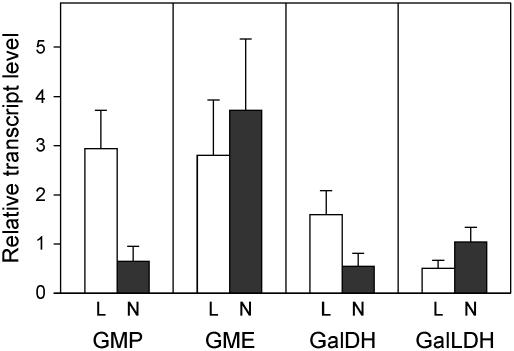
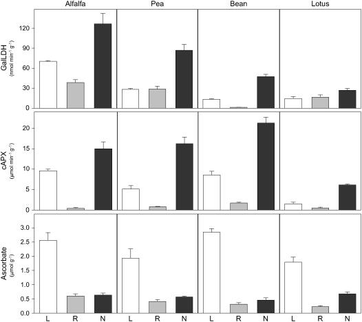
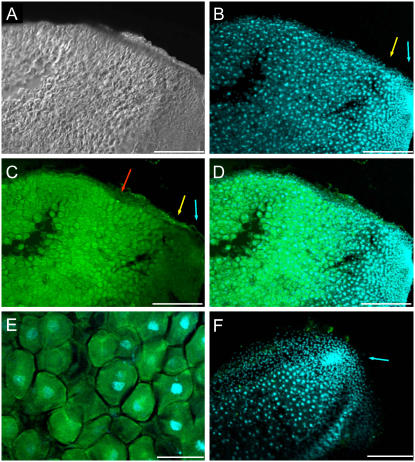
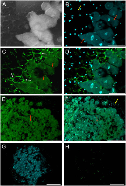
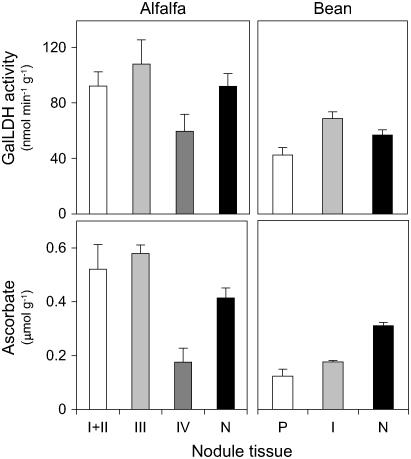
Ascorbic acid (vitamin C) is a major antioxidant and redox buffer, but is also involved in other critical processes of plants. Recently, the hypothesis has been proposed that legume nodules are unable to synthesize ascorbate and have to import it from the shoot or root, thus providing a means by which the plant regulates nodule senescence. The last step of ascorbate biosynthesis in plants is catalyzed by L-galactono-1,4-lactone dehydrogenase (GalLDH). The mRNAs encoding GalLDH and three other enzymes involved in ascorbate biosynthesis are clearly detectable in nodules. Furthermore, an active membrane-bound GalLDH enzyme is present in nodule mitochondria. Biochemical assays on dissected nodules reveal that GalLDH activity and ascorbate are correlated in nodule tissues and predominantly localized in the infected zone, with lower levels of both parameters (relative to the infected tissues) in the apex (87%) and senescent region (43%) of indeterminate nodules and in the peripheral tissues (65%) of determinate nodules. In situ RNA hybridization showed that the GalLDH mRNA is particularly abundant in the infected zone of indeterminate and determinate nodules. Thus, our results refute the hypothesis that ascorbate is not synthesized in nodules and lend support to a previous conclusion that ascorbate in the infected zone is primarily involved in the protection of host cells against peroxide damage. Likewise, the high ascorbate and GalLDH activity levels found in the apex of indeterminate nodules strongly suggest a participation of ascorbate in additional functions during symbiosis, possibly related to cell growth and division and to molecular signaling.


Figures

Figure 1.
Figure 1.
Structure of the GalLDH gene of Lotus. Comparison of the GalLDH genes of Lotus (LjGalLDH), Arabidopsis (AtGalLDH, locus At3g47930), and Oryza sativa (OsGalLDH1, locus Os11g04740; OsGalLDH2, locus Os12g04520). Lengths of exons (open reading frames, black boxes; UTRs, gray boxes) and introns (white boxes) are given in base pairs and are drawn to scale, except for introns 2 and 3 of LjGalLDH.
Figure 2.
Figure 2.
Expression of four genes involved in the d-Man/l-Gal pathway for ascorbate biosynthesis in Lotus. The steady-state mRNA levels of GMP, GME, GalDH, and GalLDH were quantified in leaves, roots, and nodules by qRT-PCR. Values of leaves (L) and nodules (N) were expressed relative to roots of the same plants (which were arbitrarily given a value of 1) and are means ± se of four to five different extracts from at least two series of plants grown independently.
Figure 3.
Figure 3.
Distribution of GalLDH activity, cAPX activity, and ascorbate in legumes. Leaves (L), roots (R), and nodules (N) of alfalfa and pea (indeterminate nodulation) and Lotus and bean (determinate nodulation) were simultaneously harvested at the plant ages indicated under “Materials and Methods.” Values are means ± se of three to eight different extracts from at least two series of plants grown independently. Units of enzyme activity and ascorbate content are per gram of fresh weight.
Figure 4.
Figure 4.
Localization of GalLDH mRNA in alfalfa (indeterminate) nodules. A, DIC image of a longitudinal section of a nodule. B, DAPI staining of nuclei (blue fluorescence) in the same nodule section as A. Note the high nuclei density in the meristematic zone (I; blue arrow) and invasion zone (II; yellow arrow). C, FISH of A with GalLDH antisense probe (green fluorescence). Note the very intense fluorescence in the infected zone (III; red arrow) and much lower fluorescence in zones I (blue arrow) and II (yellow arrow). D, Merged image of DAPI and FISH fluorescence signals. E, High magnification of the infected region of the nodule, showing an overlay of DAPI and FISH signals. Transcript labeling (green) is mostly observed in the host cell cytoplasm. F, Overlaid images of a nodule longitudinal section with DAPI and FISH using a GalLDH sense probe (control). Note the virtual absence of green fluorescence as well as the very intense labeling of the nuclei in the meristem zone (I; blue arrow). Magnification bars = 300 μm in A to D and F; 75 μm in E.
Figure 5.
Figure 5.
Localization of GalLDH mRNA in Lotus (determinate) nodules. A, DIC image showing the peripheral tissues and infected region of a nodule. Note the large polygonal, infected cells. B, DAPI staining (blue fluorescence) of the same nodule section as A. The nuclei of infected cells (red arrows) are larger than those of cortical cells (yellow arrow) and are localized toward the center of the cells. C, FISH signal in the same nodule section as A with GalLDH antisense probe. The green fluorescence is localized in the polygonal, large infected cells, with poor signal in the peripheral tissue. Note also the bright green autofluorescence of cell walls at the peripheral cell layers adjacent to infected cells (white arrows). Autofluoresecence was often observed at high magnifications but can be easily distinguished from the FISH fluorescence signal of the host cell cytoplasm (red arrows). D, Merged image of DAPI and FISH fluorescence signals. E, FISH signal in the central, infected region of nodules (red arrows). F, Merged image of DAPI and FISH signals. Note the labeling of nuclei in the cortical cells (yellow arrow), which display a low fluorescence signal compared with the infected cells (red arrows). G, Low magnification view of a nodule after DAPI labeling. H, The same nodule section as G incubated with a GalLDH sense probe as a negative control. Note the virtual absence of green fluorescence signal. Magnification bars = 75 μm in A to D; 300 μm in E to H.
Figure 6.
Figure 6.
Distribution of GalLDH activity and ascorbate in tissues of alfalfa (indeterminate) and bean (determinate) nodules. Both parameters were measured in zones I + II (meristem + invasion), III (infected), and IV (senescent) of alfalfa nodules, and in the peripheral tissues (P) and infected zone (I) of bean nodules. Whole nodules (N) were extracted in parallel as controls for recovery of GalLDH activity and ascorbate. Values are means ± se of three to five different extracts from nodules of at least two series of plants grown independently. Units of GalLDH activity and ascorbate content are per gram of fresh weight.

References

    1. Alesandrini F, Mathis R, Van de Sype G, Hérouart D, Puppo A (2003) Possible roles for a cysteine protease and hydrogen peroxide in soybean nodule development and senescence. New Phytol 158: 131–138
    1. Arrigoni O, Arrigoni-Liso R, Calabrese G (1977) Ascorbic acid requirement for biosynthesis of hydroxyproline-containing proteins in plants. FEBS Lett 82: 135–138 - PubMed
    1. Arrigoni O, De Tullio MC (2002) Ascorbic acid: much more than an antioxidant. Biochim Biophys Acta 1569: 1–9 - PubMed
    1. Bartoli CG, Guiamet JJ, Kiddle G, Pastori GM, Di Cagno R, Theodoulou FL, Foyer CH (2005) Ascorbate content of wheat leaves is not determined by maximal L-galactono-1,4-lactone dehydrogenase (GalLDH) activity under drought stress. Plant Cell Environ 28: 1073–1081
    1. Bartoli CG, Pastori GM, Foyer CH (2000) Ascorbate biosynthesis in mitochondria is linked to the electron transport chain between complexes III and IV. Plant Physiol 123: 335–343 - PMC - PubMed

Publication types

MeSH terms