. 2006 Aug;141(4):1293-305.
doi: 10.1104/pp.106.081406. Epub 2006 Jun 9.

Analysis of the subcellular localization, function, and proteolytic control of the Arabidopsis cyclin-dependent kinase inhibitor ICK1/KRP1

Affiliations

Analysis of the subcellular localization, function, and proteolytic control of the Arabidopsis cyclin-dependent kinase inhibitor ICK1/KRP1

Marc J Jakoby et al. Plant Physiol. 2006 Aug.

Abstract

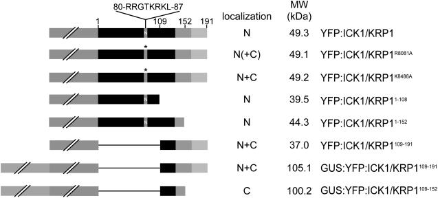
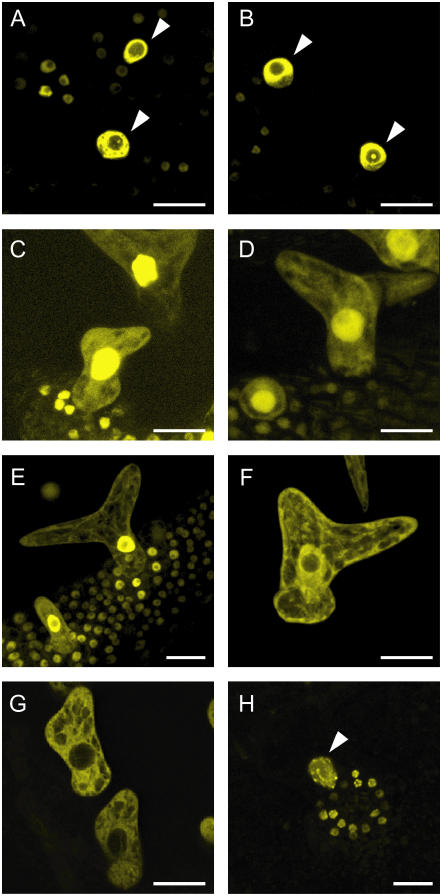
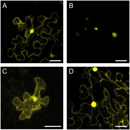
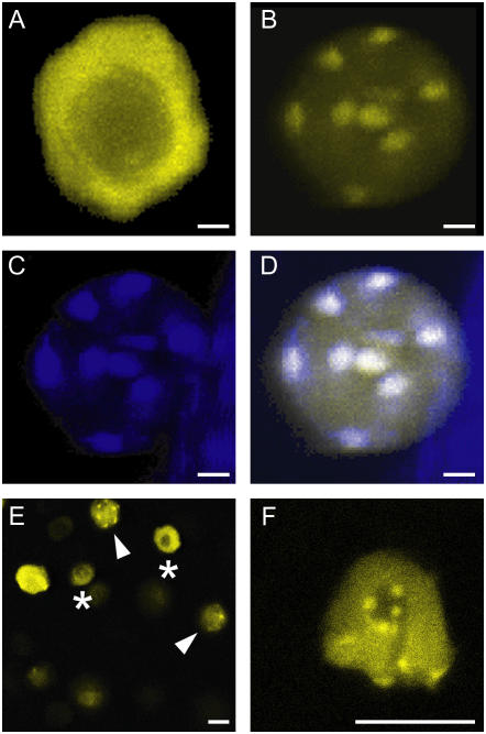
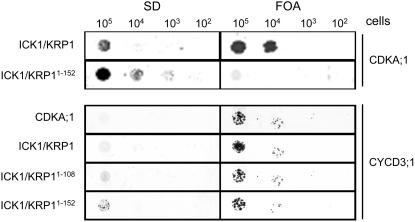
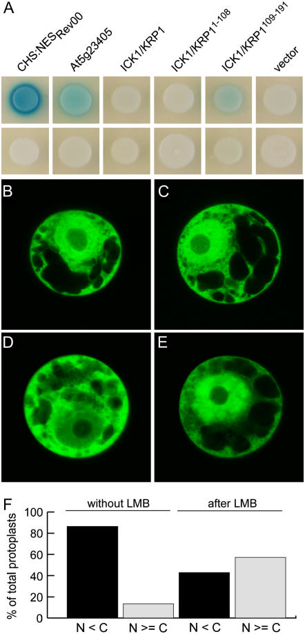
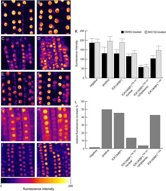
Recent studies have shown that cyclin-dependent kinase (CDK) inhibitors can have a tremendous impact on cell cycle progression in plants. In animals, CDK inhibitors are tightly regulated, especially by posttranslational mechanisms of which control of nuclear access and regulation of protein turnover are particularly important. Here we address the posttranslational regulation of INHIBITOR/INTERACTOR OF CDK 1 (ICK1)/KIP RELATED PROTEIN 1 (KRP1), an Arabidopsis (Arabidopsis thaliana) CDK inhibitor. We show that ICK1/KRP1 exerts its function in the nucleus and its presence in the nucleus is controlled by multiple nuclear localization signals as well as by nuclear export. In addition, we show that ICK1/KRP1 localizes to different subnuclear domains, i.e. in the nucleoplasm and to the chromocenters, hinting at specific actions within the nuclear compartment. Localization to the chromocenters is mediated by an N-terminal domain, in addition we find that this domain may be involved in cyclin binding. Further we demonstrate that ICK1/KRP1 is an unstable protein and degraded by the 26S proteasome in the nucleus. This degradation is mediated by at least two domains indicating the presence of at least two different pathways impinging on ICK1/KRP1 protein stability.


Figures

Figure 1.
Figure 1.
Overview of ICK1/KRP1 constructs analyzed in planta. ICK1/KRP1 was fused to the C terminus of YFP or to a GUS-YFP fusion (both reporters represented by a gray box with two slashes). The position of the NLS from amino acids R80 to L87 is marked with N and the amino acid sequence is given on top. The mutated NLS is marked by an asterisk. The cyclin- and CDK-binding sites at the C terminus of ICK1/KRP1 are highlighted by a medium and light gray box, respectively. The subcellular localization of the fusion constructs is given as nuclear (N) and/or cytoplasmic (C), parentheses indicate weak localization.
Figure 2.
Figure 2.
Subcellular localization of ICK1/KRP1 in trichomes (A–H). Confocal laser-scanning micrographs of Arabidopsis trichomes on young rosette leaves. A, In plants expressing YFP:ICK1/KRP1 under control of the GL2 promoter the YFP signal is only detectable in the nucleus. B, Most plants expressing the NLS mutant version YFP:ICK1/KRP1R80/81A show only nuclear fluorescence. C, In a few transgenic lines a weak YFP signal can be also detected in the cytoplasm by increasing the gain setting of the detector. D, All transgenic lines expressing YFP:ICK1/KRP1K84/86A show a YFP signal in the cytoplasm as well as in the nucleus. E and F, A nuclear and cytoplasmic localization of YFP fluorescence can be seen in plants expressing ProGL2:YFP:ICK1/KRP1109191 and ProGL2:GUS:YFP:ICK1/KRP1109191, respectively. G, In plants expressing ProGL2:GUS:YFP:ICK1/KRP1109152 the YFP signal is excluded from the nucleus. H, Misexpression of YFP:ICK1/KRP11108 results in an exclusively nuclear localization; note the patchy appearance of the YFP signal in the nuclei of trichomes and the surrounding socket cells. Arrowheads in A, B, and H indicate trichome nuclei. Scale bars in A to H represent 20 μm.
Figure 3.
Figure 3.
Subcellular interaction of ICK1/KRP1 with CDKA;1 (A–D). Confocal laser-scanning micrographs of the abaxial surface of Nicotiana benthamiana leaves. A, Pro35S:CDKA;1:YFP transient expression in a N. benthamiana leaf. The YFP signal can be detected in the cytoplasm and the nucleus. B, BiFC signal of CDKA;1 and the full-length ICK1/KRP1 fluorescence can be detected exclusively in the nucleus. C, BiFC signal of CDKA;1 and ICK1/KRP1109–191 can be detected in the cytoplasm and the nucleus. D, BiFC signal of CDKA;1 and ICK1/KRP1K84/86A fluorescence can be detected in the cytoplasm and the nucleus. Scale bars in A to D represent 40 μm.
Figure 4.
Figure 4.
Subnuclear localization of ICK1/KRP1 (A–F). Confocal laser-scanning micrographs of plants expressing ProGL2:YFP:ICK1/KRP1 (A, E, and F) and ProGL2:YFP:ICK1/KRP11108 (B–D). A, Apart from the nucleolus the fluorescence of YFP:ICK1/KRP1 is evenly distributed in trichome nuclei. B, In contrast, YFP:ICK1/KRP11108 localizes to some patches in trichome nuclei. C, Staining of the same nucleus shown in B with DAPI; the bright spots are the chromocenters. D, Overlay of the YFP and DAPI signals from B and C, demonstrating that YFP:ICK1/KRP11108 localizes to chromocenters in trichome nuclei. E, Epidermis of a root expressing ProGL2:YFP:ICK1/KRP1 containing nuclei with different fluorescence patterns; asterisks indicate nuclei with homogenous fluorescence as seen in A, arrowheads indicate nuclei with a punctuate appearance as seen in B. F, Close up of a nucleus of a root expressing ProGL2:YFP:ICK1/KRP1 with a punctuate fluorescence pattern. Scale bars in A to F represent 10 μm.
Figure 5.
Figure 5.
Interaction of ICK1/KRP1 with CYCD3;1. The left column shows yeast cells growing on SD medium without uracil, the right column shows yeast cells on SD media plates containing 5-FOA (FOA). Protein-protein interaction results in growth on FOA but not on SD plates; no interaction results in growth on SD but not on FOA. All yeast lines grew on SD with uracil (data not shown). Prey vectors (pNui) are listed on the left, bait vectors (pMet) on the right. CDKA;1 interacts with ICK1/KRP1 but not with ICK1/KRP11–152. In contrast, CYCD3;1 interacts with all three ICK1/KRP1 variants, full-length ICK1/KRP1, ICK1/KRP11–152, and ICK1/KRP11–108.
Figure 6.
Figure 6.
Interaction of ICK1/KRP1 with the nuclear export machinery (A). Yeast strain EGY48[p8op-lacZ] was cotransformed with pGilda bait and pB42AD prey plasmids as indicated and transformed yeast cells were replated on indicator plates containing Gal and X-gal to assay protein interactions. Top row, yeast colonies containing XPO1 in the bait vector and the indicated prey constructs; bottom row, yeast colonies transformed with an empty bait vector and the indicated prey constructs. The two positive controls, a fragment of the HIV Rev protein containing the NES (NESRev) and At5g23405, displayed a strong and medium-strong interaction with XPO1, respectively. Whereas ICK1/KRP1 and ICK1/KRP11–108 did not interact with XPO1, a weak but significant and reproducible interaction of XPO1 with ICK1/KRP1109–191 was detected. B to F, Nuclear export by XPO1 was assayed in tobacco Bright-Yellow 2 protoplasts using the inhibitor LMB. B and C, Protoplasts transfected with either YFP or GUS:YFP:ICK1/KRP1109191 (D and E) were incubated with ethanol (B and D; 0.7% final concentration) or with LMB (C and E; 0.5 μm in ethanol) for 1 h and analyzed for YFP fluorescence using confocal laser-scanning microscopy. While YFP localization did not change during the treatment, the localization of GUS:YFP:ICK1/KRP1109–191 changed to a more nuclear localization after incubation with LMB. F, Protoplasts producing GUS:YFP:ICK1/KRP1109–191 were analyzed for the percentage of those that showed equal or more YFP fluorescence in the nucleus than in the cytoplasm without (8 out of 59) or with LMB treatment (36 out of 63).
Figure 7.
Figure 7.
Degradation of ICK1/KRP1 in nuclei (A–J). False color confocal laser-scanning micrographs in false colors of Arabidopsis roots; color scale giving at the bottom. Left column shows roots treated with DMSO only, right column displays roots treated with MG132 in DMSO. A and B, In the negative control, NLS:GFP:GUS expressed from the GL2 promoter shows no altered subcellular distribution or increased fluorescence when treated with DMSO, n = 142 (A) in comparison to treatment with MG132, n = 142 (B). C and D, The positive control, YFP:DB expressed from the CDKA;1 promoter, strongly accumulates after treatment with MG132, nDMSO = 117 cells and nMG132 = 113 cells. E and F, YFP:ICK1/KRP1 accumulates in nuclei after treatment with MG132, nDMSO = 334 cells and nMG132 = 439 cells; no accumulation in the cytoplasm was found. G and H, YFP:ICK1/KRP1109–191 displays a slight accumulation of nuclear fluorescence, nDMSO = 216 cells and nMG132 = 235 cells; no cytoplasmic accumulation could be observed, nDMSO = 179 cells and nMG132 = 189 cells. I and J, The N terminus of ICK1/KRP1, ICK1/KRP11–108, strongly accumulates after treatment with MG132, nDMSO = 614 cells and nMG132 = 645 cells. K, Quantification of nuclear and, if applicable, cytoplasmic fluorescence of the above shown untreated (DMSO only) or treated (MG132 in DMSO) roots. Student's t tests were performed to analyze the statistical significance of the observed distributions: No statistical significant change was found for the DMSO-treated versus MG132-treated negative control (P = 0.7285). Also, no significant difference could be found for the fluorescence in the cytoplasm of the solvent- versus the MG132-treated YFP:ICK1/KRP1109–191 lines (P = 0.7957). For all other genotypes a statistically significant difference could be found between the DMSO and the MG132 treatments (P ≤ 0.001). L, Normalization of the fluorescent intensities shown in K; the fluorescence intensities observed with the DMSO treatment were set to 100% and the relative increase by the MG132 treatment is given in percentages. For ICK1/KRP1, 439 nuclei of MG132-treated roots and 334 nuclei of DMSO-treated roots were analyzed. For ICK1/KRP1109191, the nuclear fluorescence of 235 nuclei of MG132-treated and 216 nuclei of DMSO-treated roots and the cytoplasm of 189 MG132- and 179 DMSO-treated cells was quantified. For ICK1/KRP11108, 645 nuclei of MG132- and 614 nuclei of DMSO-treated roots were analyzed. For the negative control 142 nuclei of MG132- and 142 nuclei of DMSO-treated roots were analyzed. For the positive control 113 nuclei of MG132- and 117 nuclei of DMSO-treated plants were analyzed.

References

    1. Bisbis B, Delmas F, Joubes J, Sicard A, Hernould M, Inze D, Mouras A, Chevalier C (2006) Cyclin-dependent kinase inhibitors regulate the CDK/cyclin complex activities in endoreduplicating cells of developing tomato fruit. J Biol Chem 281: 7374–7383 - PubMed
    1. Carrano AC, Eytan E, Hershko A, Pagano M (1999) SKP2 is required for ubiquitin-mediated degradation of the CDK inhibitor p27. Nat Cell Biol 1: 193–199 - PubMed
    1. Chan GK, Liu ST, Yen TJ (2005) Kinetochore structure and function. Trends Cell Biol 15: 589–598 - PubMed
    1. Clough SJ, Bent AF (1998) Floral dip: a simplified method for Agrobacterium-mediated transformation of Arabidopsis thaliana. Plant J 16: 735–743 - PubMed
    1. Coelho CM, Dante RA, Sabelli PA, Sun Y, Dilkes BP, Gordon-Kamm WJ, Larkins BA (2005) Cyclin-dependent kinase inhibitors in maize endosperm and their potential role in endoreduplication. Plant Physiol 138: 2323–2336 - PMC - PubMed

Publication types

MeSH terms

Substances