. 2006 Aug;141(4):1316-27.
doi: 10.1104/pp.106.079327. Epub 2006 Jun 9.

Identification of features regulating OST1 kinase activity and OST1 function in guard cells

Affiliations

Identification of features regulating OST1 kinase activity and OST1 function in guard cells

Christophe Belin et al. Plant Physiol. 2006 Aug.

Abstract

The phytohormone abscisic acid (ABA) mediates drought responses in plants and, in particular, triggers stomatal closure. Snf1-related kinase 2 (SnRK2) proteins from several plant species have been implicated in ABA-signaling pathways. In Arabidopsis (Arabidopsis thaliana) guard cells, OPEN STOMATA 1 (OST1)/SRK2E/SnRK2-6 is a critical positive regulator of ABA signal transduction. A better understanding of the mechanisms responsible for SnRK2 protein kinase activation is thus a major goal toward understanding ABA signal transduction. Here, we report successful purification of OST1 produced in Escherichia coli: The protein is active and autophosphorylates. Using mass spectrometry, we identified five target residues of autophosphorylation in recombinant OST1. Sequence analysis delineates two conserved boxes located in the carboxy-terminal moiety of OST1 after the catalytic domain: the SnRK2-specific box (glutamine-303 to proline-318) and the ABA-specific box (leucine-333 to methionine-362). Site-directed mutagenesis and serial deletions reveal that serine (Ser)-175 in the activation loop and the SnRK2-specific box are critical for the activity of recombinant OST1 kinase. Targeted expression of variants of OST1 kinase in guard cells uncovered additional features that are critical for OST1 function in ABA signaling, although not required for OST1 kinase activity: Ser-7, Ser-18, and Ser-29 and the ABA-specific box. Ser-7, Ser-18, Ser-29, and Ser-43 represent putative targets for regulatory phosphorylation and the ABA-specific box may be a target for the binding of signaling partners in guard cells.


Figures
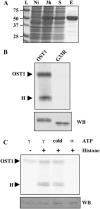
Figure 1.
Figure 1.
OST1 recombinant protein is active and autophosphorylates. A, Different steps of 10xHis-OST1 production and native purification, SDS-PAGE, and Coomassie staining. L, Ladder; Ni, noninduced bacteria lysate; 3 h, bacteria lysate 3 h after isopropylthio-β-galactoside induction; S, soluble fraction; E, elution. B, In vitro kinase activity (top) of the native recombinant 10xHis-OST1 (OST1) and the mutated version 10xHis-OST1G33R (G33R). C, In vitro kinase activity (top) of the native recombinant 10xHis-OST1 without (−) or with (+) histone III-S substrate and with usual [γ-33P]ATP (γ), [α-33P]ATP (α), or [γ-33P]ATP followed by a pulse of cold ATP 1 min before stopping the reaction (cold). B and C, Arrowheads indicate autophosphorylation (OST1) and phosphorylation of histone III-S substrate (H). The bottom image shows the protein level by immunodetection using antibodies directed against the poly-His tag (WB).
Figure 2.
Figure 2.
In planta OST1 phosphorylation is required for its activity. Kinase assay of proteins immunoprecipitated from GCP extracts using a serum that specifically recognizes OST1. Protoplasts were treated for 30 min with 30 μm ABA (+) or with ethanol solvent (−). Immunoprecipitated proteins were treated with a calf intestinal phosphatase (+CIP) or were submitted to the same treatment without any phosphatase (−CIP). The faint band at high Mr corresponds to autophosphorylation and the high-intensity band at low Mr represents histone phosphorylation.
Figure 3.
Figure 3.
Phosphorylated residues identified on recombinant OST1 by MS. Arrows indicate positions of identified phosphorylated residues in OST1 sequence. The catalytic domain and carboxy-terminal region are represented by bold and italic letters, respectively. Shaded sequences are not covered by MS analysis. Underlined stretches represent a conserved ATP-binding site GXGXXG (ATP) and activation segment (T loop). The black circle shows Gly-33, which results in an inactive protein when mutated to Arg.
Figure 4.
Figure 4.
Phosphorylation of Ser-175 is required for OST1 kinase activity. A, Point mutations on Ser-7, Ser-18, Ser-29, and Ser-43. B, Point mutations on Ser-175 and Thr-176. A and B, Each residue was mutated to Ala or Asp. As in Figure 1, the top band corresponds to autophosphorylation (OST1) and the bottom band corresponds to phosphorylation of histone III-S (H). The bottom sections (WB) show the protein level by immunodetection using antibodies directed against the poly-His tag.
Figure 5.
Figure 5.
OST1 expression is targeted to guard cells where it is activated by ABA and limits transpiration. A, Histochemical detection of GUS activity in the leaf of a representative line srk2e/PROOST1:3xHA-GUS. s, Stomata; v, vascular tissue. B, In-gel kinase assay of guard cell total protein extracts with histone III-S as the substrate. The arrowhead indicates the 42-kD band corresponding to OST1. C, Infrared thermography false color picture of detached leaves from Col and srk2e plants. The histogram represents the quantification of this picture (mean ± sd).
Figure 6.
Figure 6.
Several phosphorylable Ser residues are required for OST1 function in planta. A, In vitro biochemical activity of recombinant proteins produced in E. coli: quantification of one to four kinase assays as shown in Figure 4 (0 = no activity; dotted line = OST1 activity). B, Quantification of infrared thermography images of detached leaves. Black, white, and gray bars, respectively, represent wild-type (Col-0), mutant (srk2e), and one representative line srk2e/PROOST1:3xHA-X (where X = uidA, OST1, or a point-mutated version of OST1) for each construct (mean ± sd, n = 3 or 4 leaves). C, In planta activation of fusion proteins in response to ABA. In vitro kinase assay on proteins immunoprecipitated from plants treated (+) or not (−) by 30 μm ABA for 3 h. Values represent normalized activity of each version: quantification of the gel using ImageJ, subtraction of the background (srk2e+ = 0), and values normalized (OST1+ = 1).
Figure 7.
Figure 7.
SnRK2 C terminus contains two conserved boxes. Alignment of the C-terminal regions of the 10 Arabidopsis SnRK2s. Boxes show two conserved features: the SnRK2-specific box, which is conserved among all 10 kinases, and the ABA-specific box only found in strongly ABA-responsive kinases. Arrows indicate the end of the five truncated versions we have generated.
Figure 8.
Figure 8.
Truncation of the SnRK2-specific feature abolishes recombinant kinase activity in vitro. The top band corresponds to autophosphorylation and the bottom one to phosphorylation of histone III-S (H). The bottom image indicates protein level by immunoblot against the His tag (WB). OST1, Full-length recombinant protein. Δ348, Δ331, Δ320, Δ302, and Δ280 refer to the position of the last residue of each truncated version.
Figure 9.
Figure 9.
Impact of C-terminal deletions on OST1 in planta activity. A, In vitro biochemical activity of recombinant proteins produced in E. coli: quantification of one to three kinase assays as shown in Figure 8 (0 = no detectable activity; dotted line = OST1 activity). B, Histograms represent quantification of infrared thermography pictures of detached leaves. Black, white, and gray bars, respectively, represent the wild type (Col-0), mutant (srk2e), and one representative line srk2e/PROOST1:3xHA-X (where X = OST1 or a truncated version of OST1) for each construct (mean ± sd, n = 3 or 4 leaves). C, In planta activation of fusion proteins in response to ABA. In vitro kinase assay on proteins immunoprecipitated from plants treated (+) or not (−) by 30 μm ABA for 3 h. Values represent normalized activity of each version: quantification of the gel using ImageJ, subtraction of the background (srk2e+ = 0), and values normalized (OST1+ = 1).
Figure 10.
Figure 10.
The ABA box is required for OST1 function in response to ABA in guard cells. Stomatal aperture in darkness (black bars) and after 3 h of light without (white bars) or with 10 μm ABA (hatched bars) in the incubation medium (mean ± sem; n > 120 stomata). One representative line is shown for each of the constructs: srk2e, srk2e complemented by the 3xHA-OST1 construct (OST1), and srk2e expressing either 3xHA-OST1Δ331 (Δ331) or 3xHA-OST1Δ348 (Δ348) fusion proteins.

References

    1. Adams JA (2003) Activation loop phosphorylation and catalysis in protein kinases: Is there functional evidence for the autoinhibitor model? Biochemistry 42: 601–607 - PubMed
    1. Anderberg RJ, Walker-Simmons MK (1992) Isolation of a wheat cDNA clone for an abscisic acid-inducible transcript with homology to protein kinases. Proc Natl Acad Sci USA 89: 10183–10187 - PMC - PubMed
    1. Bensmihen S, To A, Lambert G, Kroj T, Giraudat J, Parcy F (2004) Analysis of an activated ABI5 allele using a new selection method for transgenic Arabidopsis seeds. FEBS Lett 561: 127–131 - PubMed
    1. Boudsocq M, Barbier-Brygoo H, Lauriere C (2004) Identification of nine sucrose nonfermenting 1-related protein kinases 2 activated by hyperosmotic and saline stresses in Arabidopsis thaliana. J Biol Chem 279: 41758–41766 - PubMed
    1. Droillard MJ, Thibivilliers S, Cazale AC, Barbier-Brygoo H, Lauriere C (2000) Protein kinases induced by osmotic stresses and elicitor molecules in tobacco cell suspensions: two crossroad MAP kinases and one osmoregulation-specific protein kinase. FEBS Lett 474: 217–222 - PubMed

Publication types

MeSH terms

LinkOut - more resources