. 2006 Jul;116(7):1994-2004.
doi: 10.1172/JCI27698. Epub 2006 Jun 8.

Serine protease HtrA1 modulates chemotherapy-induced cytotoxicity

Affiliations

Serine protease HtrA1 modulates chemotherapy-induced cytotoxicity

Jeremy Chien et al. J Clin Invest. 2006 Jul.

Abstract

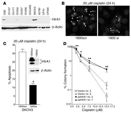
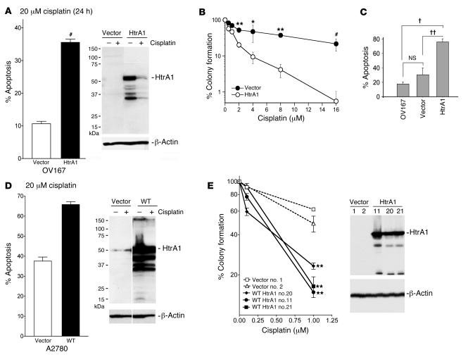
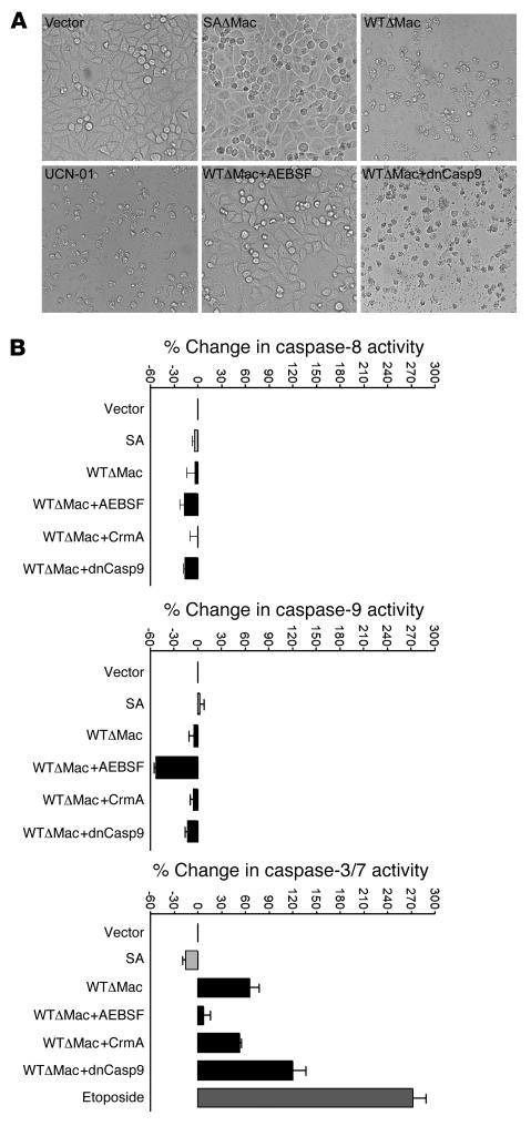
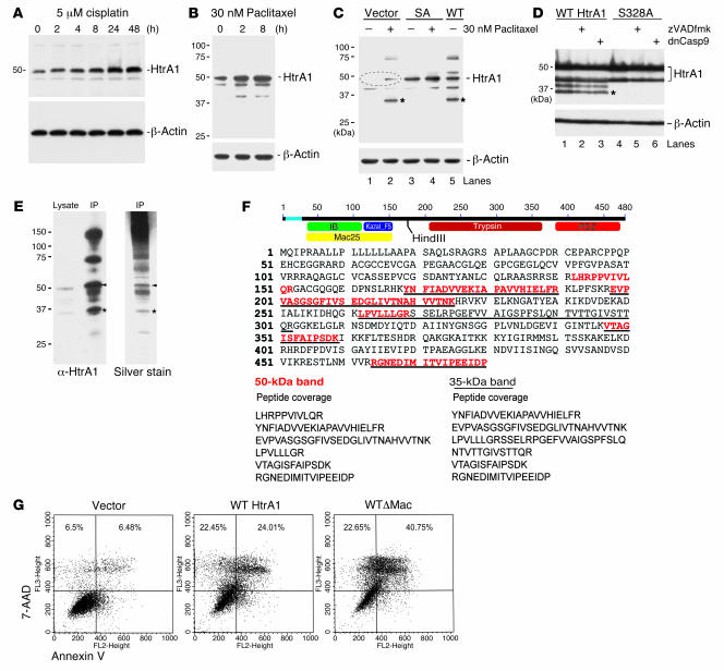
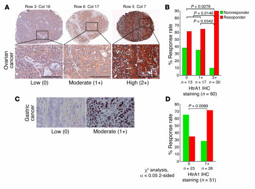
Resistance to chemotherapy presents a serious challenge in the successful treatment of various cancers and is mainly responsible for mortality associated with disseminated cancers. Here we show that expression of HtrA1, which is frequently downregulated in ovarian cancer, influences tumor response to chemotherapy by modulating chemotherapy-induced cytotoxicity. Downregulation of HtrA1 attenuated cisplatin- and paclitaxel-induced cytotoxicity, while forced expression of HtrA1 enhanced cisplatin- and paclitaxel-induced cytotoxicity. HtrA1 expression was upregulated by both cisplatin and paclitaxel treatment. This upregulation resulted in limited autoproteolysis and activation of HtrA1. Active HtrA1 induces cell death in a serine protease-dependent manner. The potential role of HtrA1 as a predictive factor of clinical response to chemotherapy was assessed in both ovarian and gastric cancer patients receiving cisplatin-based regimens. Patients with ovarian or gastric tumors expressing higher levels of HtrA1 showed a higher response rate compared with those with lower levels of HtrA1 expression. These findings uncover what we believe to be a novel pathway by which serine protease HtrA1 mediates paclitaxel- and cisplatin-induced cytotoxicity and suggest that loss of HtrA1 in ovarian and gastric cancers may contribute to in vivo chemoresistance.


Figures

Figure 1
Figure 1. Suppression of HtrA1 by siRNA attenuates cisplatin cytotoxicity.
Sensitivity to drugs was assessed by apoptosis and clonogenic survival assay in SKOV3 cells transfected with siRNAs or antisense RNA. (A) Immunoblot analysis of HtrA1 expression in ovarian cell lines. (B) SKOV3 cells were transiently transfected with siRNAs for 48 hours and subsequently treated with 20 μM cisplatin, and apoptotic cells were visualized by Hoechst staining and counted. Apoptotic cells showing chromatin condensation and fragmentation are indicated by arrowheads. (C) Analysis of apoptosis by Hoechst staining showed a significant attenuation of apoptosis in cells transfected with HtrA1 siRNA (1900si) compared with those transfected with scrambled siRNA (1900scr). Immunoblot analysis of siRNA-transfected cells indicated efficient downregulation of HtrA1 in 1900si-transfected cells (inset). (D) SKOV3 clonal lines stably expressing antisense RNA or empty vector were treated with various concentrations of cisplatin for 24 hours, washed and grown in fresh drug-free medium for 2 weeks, stained, and counted. Stable suppression of HtrA1 promoted clonogenic survival of antisense-expressing clones (asHtrA1 no. 5 and no. 7) compared with vector-expressing clones (Vector no. 3 and 9). Data are expressed as mean ± SEM and represent 3 independent trials performed at least in triplicate. *P < 0.05, **P < 0.01, #P < 0.001 or as indicated; α = 0.05, unpaired 2-tailed Student’s t test for 2 groups, and ANOVA followed by Newman-Keuls test for multiple comparison.
Figure 2
Figure 2. Reexpression of HtrA1 promotes cisplatin toxicity.
(A) HtrA1-transfected OV167 cells showed a significant increase in cisplatin-induced cell death compared with cells transfected with vector. Immunoblot analyses indicated expression of HtrA1 in cells transfected with HtrA1. β-Actin immunoblots represent loading controls. (B) OV167 cells stably expressing HtrA1 also showed a significant decrease in clonogenic survival under cisplatin treatment compared with vector-transfected cells. (C) Increased cytotoxicity following cisplatin treatment observed in OV167 cells stably expressing HtrA1 was due to an increase in apoptosis, as these cells showed significantly higher apoptosis compared with vector-transfected or parental OV167 cell lines. (D) To test whether HtrA1 expression promotes cisplatin cytotoxicity in another HtrA1-deficient cell line, A2780 cells were transiently transfected with HtrA1 and treated with cisplatin for 24 hours. HtrA1-transfected A2780 cells showed a significant increase in cisplatin-induced cell death compared with cells transfected with vector. Immunoblot analyses indicated expression of HtrA1 in cells transfected with HtrA1. (E) A2780 clonal lines stably expressing HtrA1 (HtrA1 nos. 11, 20, and 21) showed a decrease in clonogenic survival compared to vector-transfected clones (Vector nos. 1 and 2) when treated with various concentrations of cisplatin for 24 hours. Immunoblot analyses of these stable clones indicated HtrA1 expression in A2780 clones transfected with HtrA1 (nos. 11, 20, and 21). Data are expressed as mean ± SEM and represent 3 independent trials performed at least in triplicate. *P < 0.05, **P < 0.001, #P < 0.0001, P < 0.0005, ††P < 0.005; α = 0.05, unpaired 2-tailed Student’s t test for 2 groups, and ANOVA followed by Newman-Keuls test for multiple comparison.
Figure 4
Figure 4. HtrA1-induced cell death is dependent on serine protease activity.
OV202 cells were transiently transfected with various plasmids, and cell death was analyzed by morphologic examination, MTT reduction, and LDH release assays. (A) WTΔMac-transfected cells showed extensive cell death, which could be prevented by pretreatment with 50 μg/ml serine protease inhibitor AEBSF but not by cotransfection with dnCasp9. Vector- and SAΔMac-transfected cells did not show extensive cell death. 20 μM UCN-01–treated cells were used as positive controls. (B) OV202 cells transfected with various HtrA1 constructs for 8 hours were assayed for caspase activities using specific caspase substrates provided in Promega’s Caspase-Glo assay kits. In some groups, cells were cotransfected with the caspase inhibitor CrmA (cytokine response modifier A) or dnCasp9 or were preincubated with AEBSF (50 μg/ml). No significant increase in caspase-8 or -9 activity (relative to vector control) was observed following WTΔMac transfection. However, a significant increase in caspase-3/7 activity was observed with WTΔMac transfection. Caspase-3/7 activation was not blocked by CrmA or dnCasp9 but was blocked by AEBSF. 20 μM UCN-01 treatment was used as a positive control for the caspase-3/7 activity assay. Data are from experiments performed in triplicate, and error bars represent SEM.
Figure 3
Figure 3. HtrA1 is upregulated and activated during chemotherapeutic drug treatment.
(A and B) SKOV3 cells were treated with cisplatin or paclitaxel, and lysates were taken at various time points. Immunoblot analysis of HtrA1 expression in these lysates (30 μg/lane) indicated upregulation of HtrA1 by cisplatin and paclitaxel. β-Actin immunoblots in the lower panels represent loading controls. (C) Immunoblot analysis of cells treated with paclitaxel for 24 hours indicated upregulation of HtrA1 (indicated by the dashed oval) and proteolysis of HtrA1 (35-kDa product in lane 2 indicated by an asterisk) that is dependent on HtrA1 protease activity, since protease mutant SA transfectants did not produce the smaller fragment in the presence of paclitaxel (lane 4). Forced expression of WT HtrA1 also produced a similar 35-kDa product (lane 5). (D) Pretreatment with 10 μM Z-VAD-FMK or cotransfection with dnCasp9 did not prevent proteolytic processing of HtrA1 (lanes 2 and 3). However, catalytic inactivation of HtrA1 (S328A) inhibited proteolysis of HtrA1 (lanes 4–6). Autocatalytic products are indicated by an asterisk. (E) To determine the domain composition of the 35-kDa product, it was immunoprecipitated (IP) with anti-HtrA1. Immunoblot and silver stain analyses detected 35-kDa and 50-kDa bands (indicated by asterisks and arrowheads, respectively). (F) LC-MS/MS analysis of the 35-kDa band showed peptide coverage missing in the Mac25 domain (underlined) but present in the 50-kDa band (red). (G) To compare the activities of 35-kDa and 50-kDa HtrA1, plasmid constructs (full-length and ΔMac25) were transfected into OV202 cells, and apoptotic activity was analyzed by annexin V labeling. Transfection of WTΔMac induced higher cell death compared with full-length HtrA1 (WT HtrA1).
Figure 5
Figure 5. Expression of HtrA1 correlates with patient response to chemotherapy.
(A) Immunohistochemical analysis of HtrA1 expression in primary ovarian tumors on TMA showing low (0), moderate (1+), and high (2+) levels of HtrA1 staining. A small section of TMA with 3 representative HtrA1 expression levels is shown. Scale bars represent approximately 100 μm. (B) Univariate analysis of chemoresponse showing a significant association between HtrA1 expression and chemoresponse in ovarian cancer. (C) Immunohistochemical analysis of HtrA1 expression in primary gastric tumors showing low (0) and moderate (1+) levels of HtrA1 staining. Original magnification, ×250. (D) Univariate analysis of chemoresponse showing significant association between HtrA1 expression and chemoresponse in gastric cancer; 2-sided χ2 analysis, α = 0.05.

References

    1. Yeung T.K., Germond C., Chen X., Wang Z. The mode of action of taxol: apoptosis at low concentration and necrosis at high concentration. Biochem. Biophys. Res. Commun. 1999;263:398–404. - PubMed
    1. Siddik Z.H. Cisplatin: mode of cytotoxic action and molecular basis of resistance. Oncogene. 2003;22:7265–7279. - PubMed
    1. Orr G.A., Verdier-Pinard P., McDaid H., Horwitz S.B. Mechanisms of Taxol resistance related to microtubules. Oncogene. 2003;22:7280–7295. - PMC - PubMed
    1. Fraser M., et al. Chemoresistance in human ovarian cancer: the role of apoptotic regulators. Reprod. Biol. Endocrinol. 2003;1:66. - PMC - PubMed
    1. Sherman-Baust C.A., et al. Remodeling of the extracellular matrix through overexpression of collagen VI contributes to cisplatin resistance in ovarian cancer cells. Cancer Cell. 2003;3:377–386. - PubMed

Publication types

MeSH terms