. 2006 Jul;116(7):2022-32.
doi: 10.1172/JCI28423. Epub 2006 Jun 8.

Heat shock protein 60 enhances CD4+ CD25+ regulatory T cell function via innate TLR2 signaling

Affiliations

Heat shock protein 60 enhances CD4+ CD25+ regulatory T cell function via innate TLR2 signaling

Alexandra Zanin-Zhorov et al. J Clin Invest. 2006 Jul.

Retraction in

Abstract

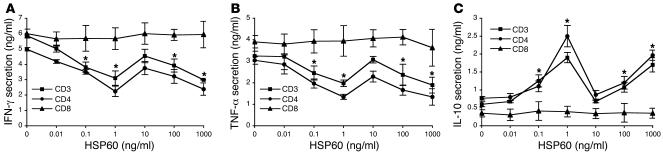
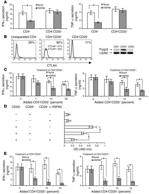
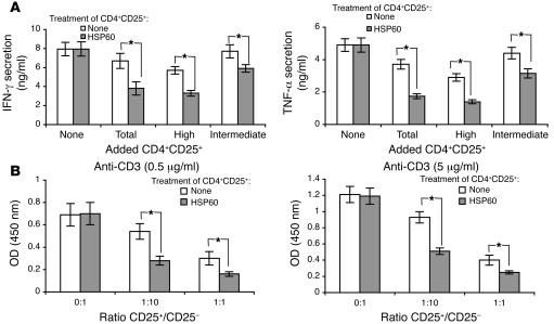
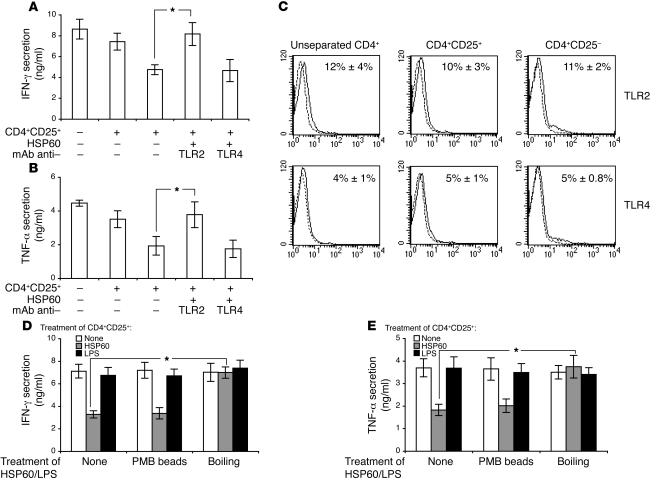
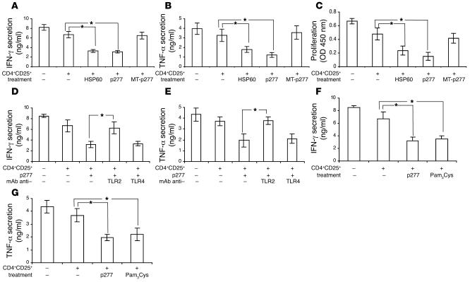
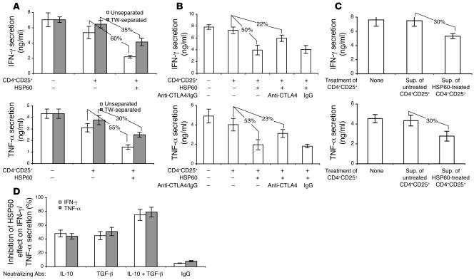
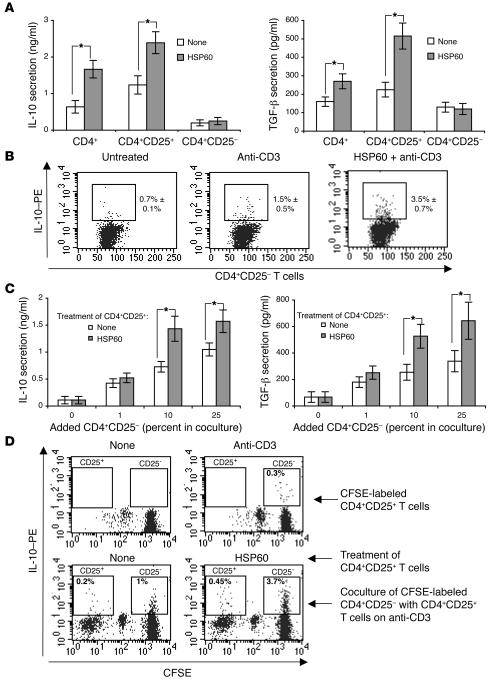
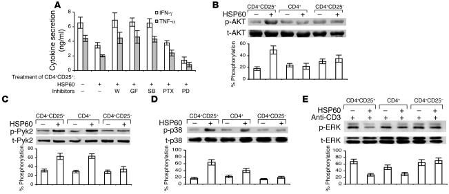
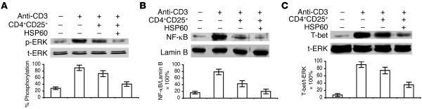
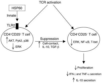
CD4+CD25+ Tregs regulate immunity, but little is known about their own regulation. We now report that the human 60-kDa heat shock protein (HSP60) acts as a costimulator of human Tregs, both CD4+CD25int and CD4+CD25hi. Treatment of Tregs with HSP60, or its peptide p277, before anti-CD3 activation significantly enhanced the ability of relatively low concentrations of the Tregs to downregulate CD4+CD25- or CD8+ target T cells, detected as inhibition of target T cell proliferation and IFN-gamma and TNF-alpha secretion. The enhancing effects of HSP60 costimulation on Tregs involved innate signaling via TLR2, led to activation of PKC, PI3K, and p38, and were further enhanced by inhibition of ERK. HSP60-treated Tregs suppressed target T cells both by cell-to-cell contact and by secretion of TGF-beta and IL-10. In addition, the expression of ERK, NF-kappaB, and T-bet by downregulated target T cells was inhibited. Thus, HSP60, a self-molecule, can downregulate adaptive immune responses by upregulating Tregs innately through TLR2 signaling.


Figures

Figure 1
Figure 1. HSP60 pretreatment inhibits anti-CD3–induced IFN-γ and TNF-α secretion and upregulates IL-10 secretion by activated CD3+ and CD4+ T cells, but not by CD8+ T cells.
Purified CD3+, CD4+, or CD8+ T cells were preincubated with the indicated concentrations of HSP60 for 2 hours, washed, and transferred to 24-well plates coated with anti-CD3 mAb (OKT; 0.5 μg/ml) in serum-free medium. The supernatants were collected after 24 hours and analyzed for IFN-γ (A), TNF-α (B), and IL-10 (C). The means ± SD of 5 different donors are shown. *P < 0.05.
Figure 2
Figure 2. The CD4+ CD25+ population mediates the inhibitory effect of HSP60 on IFN-γ and TNF-α secretion in T cells.
(A) Unseparated CD4+ and CD4+CD25+-depleted T cells were incubated with HSP60 (1 ng/ml) for 2 hours, washed, and transferred to 24-well plates coated with anti-CD3 mAb (OKT; 0.5 μg/ml) in serum-free medium. The supernatants were analyzed for IFN-γ and TNF-α. (B) Unseparated CD4+, CD4+CD25+, or CD4+CD25 T cells were checked for anti-CTLA4 and Foxp3 expression. One representative experiment of 5 is shown. (CE) CD4+CD25+ T cells were incubated with HSP60 (1 ng/ml) for 2 hours, washed, mixed in the indicated proportions with CD4+CD25 T cells (C) or with CD8+ T cells (E), and transferred to 24-well plates coated with anti-CD3 mAb (OKT; 0.5 μg/ml) in serum-free medium. The supernatants were analyzed for IFN-γ and TNF-α (C and E). Cell proliferation was determined after 96 hours at a concentration of 10% of CD4+CD25+ T cells (D). The means ± SD of 6 different donors are shown. *P < 0.05. t-ERK, total ERK.
Figure 3
Figure 3. HSP60-treated CD4+ CD25+ T cells manifest augmented regulatory effects on CD4+ CD25 T cells.
(A) Bead-sorted CD4+CD25+ T cells (total), or FACS-sorted CD4+CD25hi (high) and CD4+CD25int (intermediate) T cells, were incubated with HSP60 (1 ng/ml) for 2 hours, washed, and cocultured with CD4+CD25 T cells (ratio 1:10) on anti-CD3 in serum-free medium. The supernatants were analyzed for IFN-γ and TNF-α. The means ± SD of 5 different donors are shown. (B) CD4+CD25+ T cells were incubated with HSP60 (1 ng/ml) for 2 hours, washed, mixed in the indicated proportions with CD4+CD25 T cells, and transferred to 96-well plates coated with anti-CD3 mAb (0.5 μg/ml or 5 μg/ml). Cell proliferation was determined after 96 hours. The means ± SD of 6 different donors are shown. *P < 0.05.
Figure 4
Figure 4. The effects of HSP60 on CD4+ CD25+ T cells depend on TLR2 signaling and are not due to contaminating LPS.
(A and B) CD4+CD25+ T cells were pretreated with monoclonal anti-TLR2 or anti-TLR4 (20 μg/ml, 30 minutes). Then, the cells were incubated with HSP60 (1 ng/ml, 2 hours), washed, and cocultured with CD4+CD25 T cells (ratio 1:10) on anti-CD3 in serum-free medium. (C) The surface TLR2 or TLR4 expression on unseparated CD4+, CD4+CD25+, or CD4+CD25 T cells was determined by FACScan analysis. (D and E) CD4+CD25+ T cells were incubated (2 hours) with untreated, PMB-treated, or boiled (100°C, 30 minutes) HSP60 (1 ng/ml) or LPS (100 ng/ml). After washing, the CD4+CD25+ T cells were cocultured with test CD4+CD25 T cells (ratio 1:10) on anti-CD3 in serum-free medium. The supernatants were collected after 24 hours and analyzed for IFN-γ (A and D) and TNF-α (B and E). The means ± SD of 4 different donors are shown. *P < 0.05.
Figure 5
Figure 5. HSP60 peptide p277 or Pam3 Cys can augment the ability of CD4+ CD25+ T cells to inhibit cytokine secretion and proliferation in coculture with target CD4+ CD25 T cells via TLR2.
CD4+CD25+ T cells were incubated with HSP60, p277, MT-p277 (1 ng/ml), or Pam3Cys (100 ng/ml) for 2 hours, washed, and then cocultured with untreated CD4+CD25 T cells (ratio 1:10) on anti-CD3 in serum-free medium. The supernatants were analyzed for IFN-γ (A, D, and F) and TNF-α (B, E, and G). Proliferation was determined after 96 hours (C). In D and E, CD4+CD25+ T cells were pretreated with monoclonal anti-TLR2 or anti-TLR4 (20 μg/ml, 30 minutes). The means ± SD of 3 different donors are shown. *P < 0.05.
Figure 6
Figure 6. HSP60-induced enhancement of CD4+ CD25+ Treg function involves both contact-dependent and cytokine-dependent mechanisms.
(A) CD4+CD25+ T cells were incubated with HSP60 (1 ng/ml, 2 hours), washed, and cocultured with target CD4+CD25 T cells (ratio 1:10) on anti-CD3 in the same lower well or separately in the upper chamber of the Transwell (TW). As indicated, some CD4+CD25+ T cells were pretreated with control or monoclonal anti-CTLA4 (20 μg/ml, 30 minutes) and washed (B), or blocking anti–IL-10 and/or anti–TGF-β (10 μg/ml) mAbs (D) were added to the coculture. The supernatants were collected after 24 hours and analyzed for IFN-γ and TNF-α. The means ± SD of 5 different donors are shown. (C) CD4+CD25+ T cells were incubated with HSP60 (1 ng/ml, 2 hours), washed, and plated on anti-CD3 for 24 hours in serum-free medium. Then the supernatants (Sup.) were collected and added to test CD4+CD25 T cells in the presence of anti-CD3 mAbs. The supernatants from the CD4+CD25 T cells were collected after an additional 24 hours and analyzed for IFN-γ and TNF-α. The means ± SD of 4 different donors are shown.
Figure 7
Figure 7. HSP60 induces IL-10 and TGF-β secretion in CD4+ CD25+ T cells.
Unseparated CD4+ (A), CD4+CD25+ (AD), or CD4+CD25 T cells (A) were incubated with HSP60 (1 ng/ml) for 2 hours, washed, and transferred to 24-well plates coated with anti-CD3 mAb (OKT; 0.5 μg/ml) separately (A and B), or cocultured in the indicated proportions (C and D) in serum-free medium. IL-10 and TGF-β secretion was analyzed after 24 hours by ELISA (A and C), or by FACS (B and D). (D) CD4+CD25 T cells were labeled with CFSE and washed before coculture with CD4+CD25+ T cells. (A and C) The means ± SD of 5 different donors are shown. *P < 0.05. (B and D) FACS histograms are representative of 3 different donors. Numbers indicate average percentage ± SD of 3 different donors.
Figure 8
Figure 8. HSP60 pretreatment upregulates anti-CD3–induced phosphorylation of AKT, Pyk2, and p38 and downregulates ERK phosphorylation in CD4+ CD25+ T cells.
(A) CD4+CD25+ T cells were pretreated with 1 of the intracellular signal transduction inhibitors wortmannin (W; 5 nM), GF109203X (GF; 20 nM), SB 203580 (SB; 10 μm), pertussis toxin (PTX; 2 μg/ml), and PD 98059 (PD; 10 μm). Then, the cells were incubated with HSP60 (1 ng/ml, 2 hours), washed, and cocultured with test CD4+CD25 T cells (ratio 1:10) on anti-CD3 in serum-free medium. The supernatants were analyzed for IFN-γ and TNF-α. (BE) Unseparated CD4+, CD4+CD25+, or CD4+CD25 T cells were incubated with HSP60 (1 ng/ml) for 10 minutes and washed, and some cells (E) were exposed to anti-CD3 mAbs (60 minutes). Lysates of these cells were immunoblotted with antibodies: anti–phospho-AKT (p-AKT) and anti–total AKT (t-AKT) (B), anti–p-Pyk2 and anti–t-Pyk2 (C), anti–p-p38 and anti–t-p38 (D), or anti–p-ERK and anti–t-ERK (E). The blot of 1 experiment representative of 3 different donors is presented. Phosphorylation levels of the experiments were estimated by densitometry, and an average percentage of phosphorylation ± SD was calculated as OD of p-AKT/t-AKT, p-Pyk2/t-Pyk2, p-p38/t-p38, or p-ERK/t-ERK × 100%.
Figure 9
Figure 9. Coculture of CD4+ CD25 T cells with HSP60-treated CD4+ CD25+ T cells downregulates ERK phosphorylation and inhibits nuclear translocation of NF-κB and T-bet expression in the CD4+ CD25 T cells.
CD4+CD25 T cells were labeled with CFSE, washed, and cocultured with HSP60-treated (1 ng/ml, 2 hours) CD4+CD25+ T cells (ratio 1:10) on anti-CD3 in serum-free medium for 6 hours. The CD4+CD25 T cells were reisolated by FACS sorting, and cell lysates of these cells were immunoblotted with antibodies: anti–phospho-ERK (p-ERK) and anti–total ERK (t-ERK) (A), anti–NF-κB or anti–lamin B (B), or anti–T-bet or anti–t-ERK (C). The blot of 1 experiment representative of 4 different donors is presented. The levels of ERK phosphorylation, NF-κB, and T-bet were estimated by densitometry, and the average percentage derived from 4 different donors is shown.
Figure 10
Figure 10. Schematic diagram of the innate effects of HSP60 on Treg function.
The exposure of Tregs to HSP60 via innate TLR2 signaling amplifies their response to TCR-dependent (anti-CD3) stimulation, reflected by upregulation of AKT, Pyk2, and p38 and downregulation of ERK. These innate effects of HSP60 signaling amplify IL-10, TGF-β, and contact-dependent Treg suppressor mechanisms that impinge on TCR-activated CD4+CD25 T cells to downregulate ERK, NF-κB, and T-bet, leading, in turn, to downregulated proliferation and secretion of the proinflammatory cytokines IFN-γ and TNF-α and to upregulation of IL-10 secretion.

References

    1. Sakaguchi S., et al. Immunologic tolerance maintained by CD25+ CD4+ regulatory T cells: their common role in controlling autoimmunity, tumor immunity, and transplantation tolerance. Immunol. Rev. 2001;182:18–32. - PubMed
    1. Shevach E.M. CD4+ CD25+ suppressor T cells: more questions than answers. Nat. Rev. Immunol. 2002;2:389–400. - PubMed
    1. Thornton A.M., Piccirillo C.A., Shevach E.M. Activation requirements for the induction of CD4+CD25+ T cell suppressor function. Eur. J. Immunol. 2004;34:366–376. - PubMed
    1. Lindley S., et al. Defective suppressor function in CD4(+)CD25(+) T-cells from patients with type 1 diabetes. Diabetes. 2005;54:92–99. - PubMed
    1. Viglietta V., Baecher-Allan C., Weiner H.L., Hafler D.A. Loss of functional suppression by CD4+CD25+ regulatory T cells in patients with multiple sclerosis. J. Exp. Med. 2004;199:971–979. - PMC - PubMed

Publication types

MeSH terms