Comparative Study
. 2006 Jun 20;45(24):7415-28.
doi: 10.1021/bi060152s.

The role of protein dynamics in thymidylate synthase catalysis: variants of conserved 2'-deoxyuridine 5'-monophosphate (dUMP)-binding Tyr-261

Affiliations
Comparative Study

The role of protein dynamics in thymidylate synthase catalysis: variants of conserved 2'-deoxyuridine 5'-monophosphate (dUMP)-binding Tyr-261

Zachary Newby et al. Biochemistry. .

Abstract

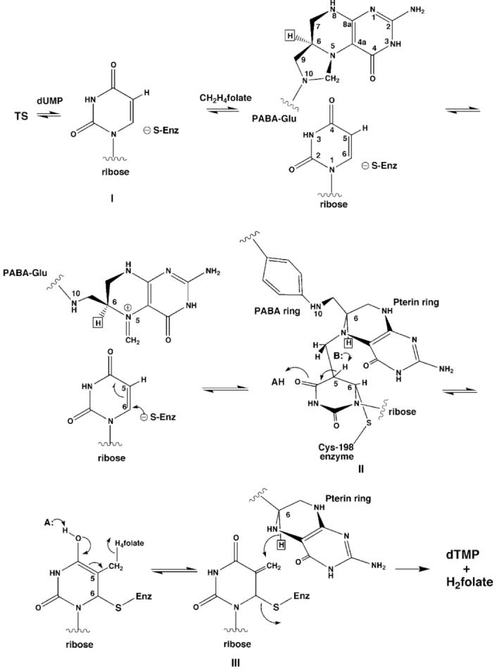
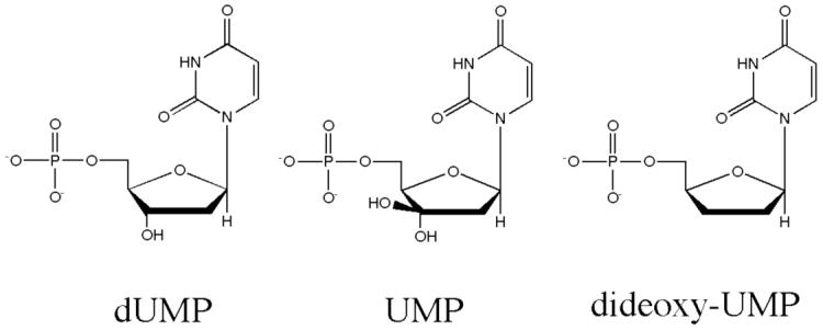
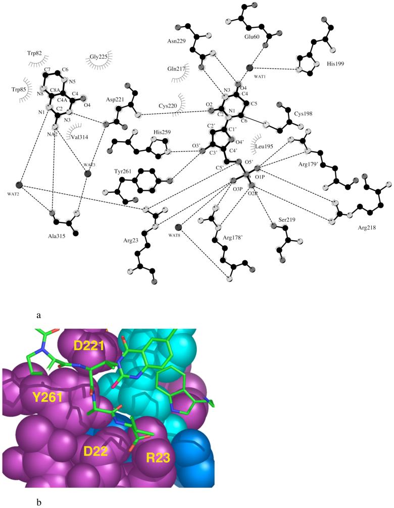
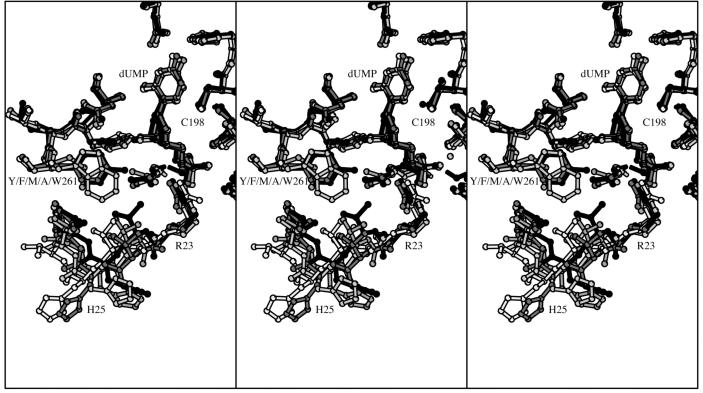
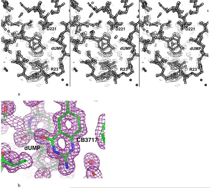
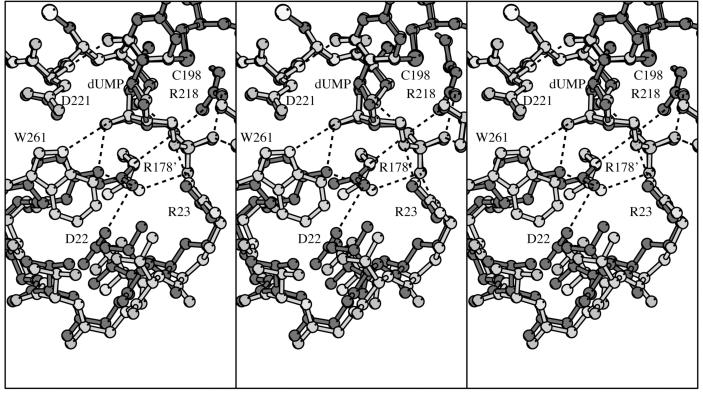
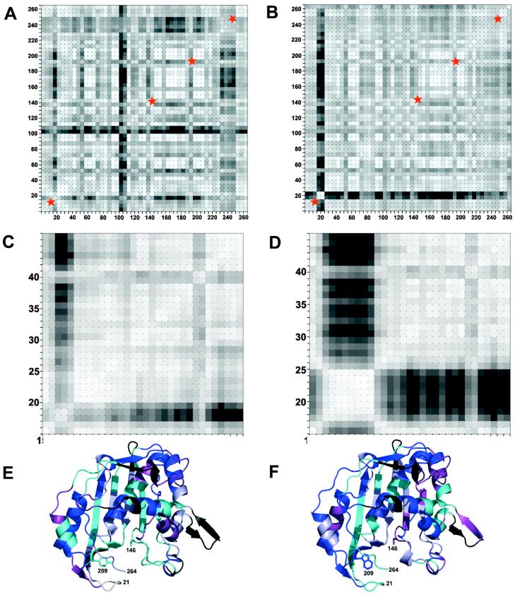
The enzyme thymidylate synthase (TS) catalyzes the reductive methylation of 2'-deoxyuridine 5'-monophosphate (dUMP) to 2'-deoxythymidine 5'-monophosphate. Using kinetic and X-ray crystallography experiments, we have examined the role of the highly conserved Tyr-261 in the catalytic mechanism of TS. While Tyr-261 is distant from the site of methyl transfer, mutants at this position show a marked decrease in enzymatic activity. Given that Tyr-261 forms a hydrogen bond with the dUMP 3'-O, we hypothesized that this interaction would be important for substrate binding, orientation, and specificity. Our results, surprisingly, show that Tyr-261 contributes little to these features of the mechanism of TS. However, the residue is part of the structural core of closed ternary complexes of TS, and conservation of the size and shape of the Tyr side chain is essential for maintaining wild-type values of kcat/Km. Moderate increases in Km values for both the substrate and cofactor upon mutation of Tyr-261 arise mainly from destabilization of the active conformation of a loop containing a dUMP-binding arginine. Besides binding dUMP, this loop has a key role in stabilizing the closed conformation of the enzyme and in shielding the active site from the bulk solvent during catalysis. Changes to atomic vibrations in crystals of a ternary complex of Escherichia coli Tyr261Trp are associated with a greater than 2000-fold drop in kcat/Km. These results underline the important contribution of dynamics to catalysis in TS.


Figures

Figure 1
Figure 1
Schematic showing the minimum mechanism for TS catalysis.
Figure 2
Figure 2
Chemical structures of dUMP, UMP and ddUMP.
Figure 3
Figure 3
a. A diagram showing dUMP and CH2H4folate interactions with EcTS. Interactions are derived from the ternary complex EcTS·5-fluoro-dUMP·CH2H4folate (52) (PDB accession code 1TSN). Carbon atoms are black while heteroatoms are dark gray (oxygen, phosphorous), light gray (nitrogen) or white (sulfur). The LcTS numbering convention is used. LcTS residue numbers, m, can be converted to EcTS residue numbers, n, by n=m-2, for m less than or equal to 89, and n=m-52 for m greater than 89. b. Slice through the active site cavity of EcTS-dUMP-CB3717 rendered as cyan space-filling atoms for dUMP, purple space-filling atoms for protein residues from the same protomer as dUMP, and blue space-filling atoms for dUMP ligands from the second protomer [R178′ (126′) and R179′ (127′)]. Y261(209) is enclosed by D221(169) above, the phosphate-binding loop below, and on either side, the C-terminal residues (shown as stick bonds) and R178′ (126′). The cofactor analog, CB3717 and Trp-85(83), in the same layer of the protein as the C-terminus, are also plotted with stick bonds. The LcTS numbering convention is used in the figure.
Figure 4
Figure 4
Stereo plots of the overlapped structures of the dUMP complexes of wild-type LcTS (black), Y261F (dark gray), Y261M (medium gray), Y261W (light gray), and Y261A (white) in the region of the dUMP binding site. The left two panels comprise a divergent-eyes stereo pair and the right two panels comprise a crossed-eyes stereo pair.
Figure 5
Figure 5
a. Stereo plot of 3σ level (Fo1-Fo2)αcalc density around the mutated residue Y261(209)W in protomer 1, where Fo1 comes from Y209W (shown in light bonds) and Fo2 is from the wild-type structure (shown in dark bonds). Positive density contours are shown with solid lines while negative density contours are shown with dashed lines. The left two panels comprise a divergent-eyes stereo pair and the right two panels comprise a crossed-eyes stereo pair. b. Density in the active site of Y209W protomer 1 from a 1.3Å (2Fo-Fc)αcalc, map contoured at 0.5 σ.
Figure 6
Figure 6
Stereo plot of EcTS Y261(209)W·dUMP·CB3717 (light gray) in the region of the mutation, overlaid with wild-type EcTS·dUMP·CB3717 (dark gray). Hydrogen bonds are shown as dashed lines. The LcTS numbering convention is used. The left two panels comprise a divergent-eyes stereo pair and the right two panels comprise a crossed-eyes stereo pair.
Figure 7
Figure 7
Grid plots showing degree of correlation in anisotropic displacement parameters between atoms identified by the (x, y) labels of the plot (EcTS numbering), for all atoms in one protomer of dimeric wild-type EcTS·dUMP·CB3717 (panel A) and Y261(209)W·dUMP·CB3717 (panel B). Correlation is indicated by similar values for the projections of the anisotropic displacement parameters along the interatomic vector (“deltas”- see text). The deltas within a small square have been averaged and the square is shaded according to the average delta value, with lighter shades indicating greater correlation. Blocks of light colored squares along the diagonal of a plot identify protein segments that potentially move as rigid bodies. Segments whose putative rigid body vibrations may be disrupted by the Y261(209) mutation are marked by red stars. Higher resolution plots of the lower left regions of (A) and (B) are shown in panels (C) and (D), respectively. These illustrate that in wild-type EcTS ternary complex, residues 22(20)-47(45) may vibrate as a rigid body, whereas in the Y261(209)W complex, resides19(17)-27(25) clearly vibrate independently of neighboring residues. The average degree of correlation of atomic B-factors (deltas) of a residue with those of its four nearest neighbors in the sequence (equivalent to the diagonal squares in panels A and B) is mapped onto a ribbon drawing of a wild-type EcTS protomer in (E) and a Y261(209)W protomer in (F), using the following color scheme: black, dark blue, purple, cyan, light blue and white, ranging in order from least to most correlated.

References

    1. Finer-Moore J, Fauman EB, Foster PG, Perry KM, Santi DV, Stroud RM. Refined structures of substrate-bound and phosphate-bound thymidylate synthase from Lactobacillus casei. J. Mol. Biol. 1993;232:1101–1116. - PubMed
    1. Aurora R, Rose GD. Seeking an ancient enzyme in Methanococcus jannaschii using ORF, a program based on predicted secondary structure comparisons. Proc Natl Acad Sci U S A. 1998;95:2818–23. - PMC - PubMed
    1. Climie S, Ruiz-Perez L, Gonzalez-Pacanowska D, Prapunwattana P, Cho SW, Stroud R, Santi DV. Saturation site-directed mutagenesis of thymidylate synthase. J. Biol. Chem. 1990;265:18776–9. - PubMed
    1. Climie S, Santi DV. Chemical synthesis of the thymidylate synthase gene. Proc. Natl. Acad. Sci., USA. 1990;87:633–637. - PMC - PubMed
    1. Climie SC, Carreras CW, Santi DV. A Complete Replacement Set of Amino Acids at the C-Terminus of Thymidylate Synthase: Quantitative Structure-Activity Relationships of Mutants of an Enzyme. Biochemistry. 1992;31:6032–6038. - PubMed

Publication types

MeSH terms

LinkOut - more resources