. 2006 Jun 12:4:17.
doi: 10.1186/1741-7007-4-17.

Neuromuscular organization and aminergic modulation of contractions in the Drosophila ovary

Affiliations

Neuromuscular organization and aminergic modulation of contractions in the Drosophila ovary

C Adam Middleton et al. BMC Biol. .

Abstract

Background: The processes by which eggs develop in the insect ovary are well characterized. Despite a large number of Drosophila mutants that cannot lay eggs, the way that the egg is moved along the reproductive tract from ovary to uterus is less well understood. We remedy this with an integrative study on the reproductive tract muscles (anatomy, innervation, contractions, aminergic modulation) in female flies.

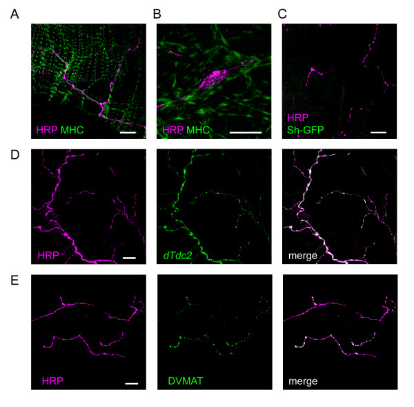
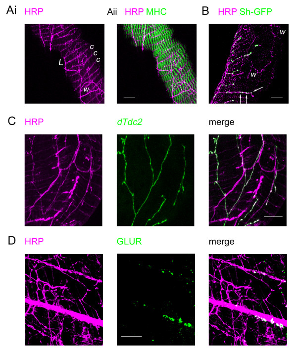
Results: Each ovary, consisting of 15-20 ovarioles, is surrounded by a contractile meshwork, the peritoneal sheath. Individual ovarioles are contained within a contractile epithelial sheath. Both sheaths contain striated muscle fibres. The oviduct and uterine walls contain a circular striated muscle layer. No longitudinal muscle fibres are seen. Neurons that innervate the peritoneal sheath and lateral oviduct have many varicosities and terminate in swellings just outside the muscles of the peritoneal sheath. They all express tyrosine decarboxylase (required for tyramine and octopamine synthesis) and Drosophila vesicular monoamine transporter (DVMAT). No fibres innervate the ovarioles. The common oviduct and uterus are innervated by two classes of neurons, one with similar morphology to those of the peritoneal sheath and another with repeated branches and axon endings similar to type I neuromuscular junctions. In isolated genital tracts from 3- and 7-day old flies, each ovariole contracts irregularly (12.5 +/- 6.4 contractions/minute; mean +/- 95% confidence interval). Peritoneal sheath contractions (5.7 +/- 1.6 contractions/minute) move over the ovary, from tip to base or vice versa, propagating down the oviduct. Rhythmical spermathecal rotations (1.5 +/- 0.29 contractions/minute) also occur. Each genital tract organ exhibits its own endogenous myogenic rhythm. The amplitude of contractions of the peritoneal sheath increase in octopamine (100 nM, 81% P < 0.02) but 1 microM tyramine has no effect. Neither affects the frequency of peritoneal sheath contractions.

Conclusion: The muscle fibres of the reproductive tract are circular and have complex bursting myogenic rhythms under octopaminergic neuromodulation. We propose a new model of tissue-specific actions of octopamine, in which strengthening of peritoneal sheath contractions, coupled with relaxation of the oviduct, eases ovulation. This model accounts for reduced ovulation in flies with mutations in the octopaminergic system.


Figures

Figure 1
Figure 1
Overview of the female reproductive tract. The innervation is outlined (magenta) from our anti-HRP staining, the muscles redrawn after [49], with only a representative set of the external uterine muscles included. The two pairs of nerves, AbNvOv (abdominal nerve to the ovary) and AbNvUt (abdominal nerve to the uterus) branch from the 5th pair of nerves emerging from the abdominal region of the CNS [13]. Scalebar: 250 μm approximately.
Figure 2
Figure 2
Ovariogram methodology. A. Ovariograms were constructed from the video micrographs as follows: a line was overlaid on the video image to cross the light/dark interface, in this case across the edge of the ovary. B. A plot of the change in position of the light dark boundary from frame to frame plotted (upper trace, 1 pixel is 1.7 μm) and of the mean square difference in intensity at each point along the line for successive pairs of frames (lower trace). The second method, recording the mean square difference in intensity provides a much greater sensitivity and also better discrimination of movement, as it takes into consideration the change in every pixel along the line, rather than just the position of the light/dark interface. Scalebar: 500 μm.
Figure 3
Figure 3
Muscle fibres of the ovary. A,B. Confocal fluorescent microscopy showing the muscle fibres of the ovariolar epithelial sheath, ES, and the ovarian peritoneal sheath, PS. The peritoneal sheath fibres overlie those of the epithelial sheath. In A, myosin (green) is Mhc-weeP26-GFP. In B, FITC-labelled anti-myosin antibody is green, with F-actin (magenta) detected with rhodamine-labelled phalloidin. Note that the ovariolar muscle fibres all circle the ovariole, whereas the peritoneal sheath muscle fibres form a meshwork across the ovarial surface. C, D. Electron micrographs of the peritoneal sheath (PS) and epithelial sheath (ES) respectively, showing longitudinal sections of sarcomeres with interdigitating thick and thin filaments (white arrow) and perforated Z-discs (black arrow). EC: egg chamber. Scalebars: A: 50 μm; B: 10 μm, C:200 nm, D:500 nm.
Figure 4
Figure 4
Muscles of the oviduct and uterus. A. Electron micrograph of a cross section of the common oviduct to show the outer muscular layer (M), surrounding the epithelial lining (EL) and the oviduct lumen (L). The epithelial lining has an apical surface rich in convoluted multimembrane structures. B. Detail of the muscle layer shows a myofibril with interdigitated thick and thin filaments (white arrow) and perforated Z-discs (black arrow). Occasionally some thick-thin filament lattice (asterisk) is seen cut transversely suggesting that not all the myofilaments run in parallel with the circular muscle layer, though these exceptions may be tangentially cut. C. A confocal image of a region of common oviduct with F-actin filaments stained (magenta) with rhodamine-phalloidin. This shows that the common oviduct is covered by circular muscle fibres (also seen in lateral oviducts, not shown); there is no evidence of longitudinal muscle fibres. Note that some of the phalloidin bands twist and a few split, and this may account for the transversely cut filaments seen in B. D. Electron micrograph of a uterus cross-section shows the muscle (M) and epithelial layers (EL). The muscle layer is considerably thicker than that of the oviduct and again exhibits perforated Z-discs. E. Confocal fluorescence image of uterine muscle, immunostained with myosin FITC-labelled antibody (green) and F-actin labelled with rhodamine-phalloidin (magenta) shows that the myofibrils within these cells seem to form side-by-side associations, often in partial register. The fibres have tapered ends. All the myofibres encircle the uterus; no longitudinal myofibres were seen. Scalebars: A: 2 μm, B: 0.5 μm, C: 20 μm, D: 2 μm, E: 10 μm.
Figure 5
Figure 5
Innervation of the ovary by a single type of aminergic fibre. A. Merged confocal images from a Wee-P26 fly of nerves immunostained with anti-HRP (magenta) and of muscle expressing MHC-GFP (green) show that the nerves run along the muscle fibres of the peritoneal sheath with varicosities at irregular intervals and at their ends. The thinner muscle fibres running vertically are part of the epithelial sheath around an ovariole. B. A similar image showing a single neural varicosity at higher magnification. C. Merged confocal images showing nerves (magenta) immunostained as above in a fly expressing CD8-GFP-Shaker (green). The absence of detectable green fluorescence indicates a lack of Type I neuromuscular junctions. D. Individual and merged confocal images showing that the expression of dTdc2-GAL4 as reported by UAS-n-syb-spH (green) co-localising (white) with nerve fibres immunostained as above (magenta). E. Individual and merged confocal images of a specimen double-labelled with anti-HRP (magenta) and an antibody to the Drosophila vesicular monoamine transporter (DVMAT, green), shows that the DVMAT staining is co-incident with neural varicosities and nerve endings with occasional smaller hot-spots along the length of the nerves. No DVMAT signal was detected in the muscle fibres. Scalebars: All 20 μm.
Figure 6
Figure 6
Innervation of the oviduct by two types of fibre. Ai. Confocal image from a Wee-P26 fly of nerves immunostained with anti-HRP (magenta) showing nerves running over the surface of the oviduct. Some nerve fibres run longitudinally (L) giving off circumferential branches (c) while others have a more random wandering path (w). Aii. The previous image merged with one of muscle expressing MHC-GFP shows that the circumferential branches run between the circular myofibrils. B. Merged confocal images of the oviduct from the same preparation as Figure 5C. The nerves (magenta) are immunostained as above in a fly expressing CD8-GFP-Shaker (green). Patches of CD8-Shaker fluorescence co-localise (white) with neural boutons (arrows) on the circumferential nerve fibres but are absent from the varicosities on the wandering fibres (w). C. Individual and merged confocal images showing that the expression of dTdc2-GAL4 as reported by UAS-n-syb-spH (green) colocalises (white) with the anti-HRP labelling (magenta) of some nerves but is absent from others. D. Individual and merged confocal images of anti-HRP labelling (magenta) and anti glutamate receptor (GLUR, green). Note that the round patches of GLUR signal are associated with the straight fibres rather than the wandering fibres. Scalebars: All 20 μm.
Figure 7
Figure 7
Independent movements in the female Drosophila reproductive tract. Ai. Simultaneous recording from 4 ovarioles, the peritoneal sheath (PS), oviduct and spermatheca, plus a stationary control. In this preparation, the bases of the two ovaries and uterus lie on the microscope slide – the spermatheca and oviducts are free to move within the saline, while the ovarioles contract within the ovary. Video recorded at 15 frames/second; only the position of the light/dark boundary is shown. Aii. Part of the record is shown expanded. Note that the spermatheca has the largest movement, when its duct uncurls for 5–6 seconds at a time. The movement in the spermatheca has a small, gradual pull on the PS and oviduct, but small, higher frequency waves are also seen in these organs. The oviduct trace shows that its contractions occur at random with respect to those of the spermatheca, sometimes occurring before, at other times during or after those of the spermatheca. Each of the ovarioles has independent activity within the ovary, ranging from fast and frequent to occasional and erratic. Note that, at the PS recording site (see Fig. 7C), the PS can be seen as a lighter colour fringe, while on the other ovary control recordings were taken where the peritoneal sheath is stretched tautly over the eggs and so no PS waves were apparent. B. Fast Fourier transforms of the data shown in A, plotting the power density from 2 to 300 contractions/minute. Although the raw FFT output extended below 2 contractions/minute, this corresponds to slow drift in the preparation rather than biological activity. Virtually no energy occurs above 60 contractions/minute (1 Hz), so recording at 2 frames/second captures the movement faithfully. Note that the ovarioles all have peaks at different frequencies. For the spermatheca, the nearly square waveform means that the main peak is accompanied by several side bands. C. The first frame of the video, showing the position at which the recordings were taken. The video is available at reduced sample rate in QuickTime format [see Additional file 1] or in other formats [50]. Scalebar: 250 μm.
Figure 8
Figure 8
Irregular wavepaths for contractions of the isolated ovary. Ai. Replicate measures of the position of the light/dark interface were made at three sites; the same colour is used for each site. For each site, three adjacent replicates were made and the signal averaged to reduce the noise. Aii. The resulting ovariogram shows that several kinds of contractions were observed, some as a single peak, others biphasic and some triphasic. Aiii. Overlaying the recordings so that waves are aligned at the tip shows that some waves move from tip to base, others from base to tip. This was observed in both the isolated ovary and the reproductive tract complete. B. Similar recordings from another preparation, showing consistent waves of contraction moving from tip to base and on into the oviduct. Scalebar: 250 μm.
Figure 9
Figure 9
An individual ovariole continues to contract in isolation. A. The first frame of the video, showing the location of the recording site. B. Contractions are monitored by changes in the mean squared difference in intensity between successive frames. Data recorded at 2 frames/second. Scalebar: 100 μm.
Figure 10
Figure 10
Octopamine, but not tyramine, increases the amplitude of peritoneal sheath contractions. Data from preparations in which the uterus and spermathecae were removed to avoid mechanical coupling. Data recorded as mean square difference in intensity, with the average height of a peak calculated for each preparation. Control: N = 41 peaks; wash N = 27; 1 μM tyramine N = 10; 10 nM octopamine N = 7. Unpaired t-tests were used to compare drug treatments, with * used to indicate P < 0.05.
Figure 11
Figure 11
There is no statistically significant change in either (A) the frequency of contraction or (B) the frequency of bursts of contractions in the peritoneal sheath with octopamine or tyramine. Same preparations and analysis as Fig. 10.

References

    1. Roeder T, Seifert M, Kahler C, Gewecke M. Tyramine and octopamine: Antagonistic modulators of behavior and metabolism. Arch Insect Biochem Physiol. 2003;54:1–13. doi: 10.1002/arch.10102. - DOI - PubMed
    1. Roeder T. Octopamine in invertebrates. Prog Neurobiol. 1999;59:533–561. doi: 10.1016/S0301-0082(99)00016-7. - DOI - PubMed
    1. Monastirioti M, Linn CE, White K. Characterization of Drosophila tyramine beta-hydroxylase gene and isolation of mutant flies lacking octopamine. J Neurosci. 1996;16:3900–3911. - PMC - PubMed
    1. Cole SH, Carney GE, McClung CA, Willard SS, Taylor BJ, Hirsh J. Two functional but noncomplementing Drosophila tyrosine decarboxylase genes: distinct roles for neural tyramine and octopamine in female fertility. J Biol Chem. 2005;280:14948–14955. doi: 10.1074/jbc.M414197200. - DOI - PubMed
    1. Kutsukake M, Komatsu A, Yamamoto D, Ishiwa-Chigusa S. A tyramine receptor gene mutation causes a defective olfactory behavior in Drosophila melanogaster. Gene. 2000;245:31–42. doi: 10.1016/S0378-1119(99)00569-7. - DOI - PubMed

Publication types

LinkOut - more resources