

Management of a patient with multiple recurrences of fibromatosis (desmoid tumor) of the breast involving the chest wall musculature
- PMID: 16768799
- PMCID: PMC1524961
- DOI: 10.1186/1477-7819-4-32
Management of a patient with multiple recurrences of fibromatosis (desmoid tumor) of the breast involving the chest wall musculature
Abstract
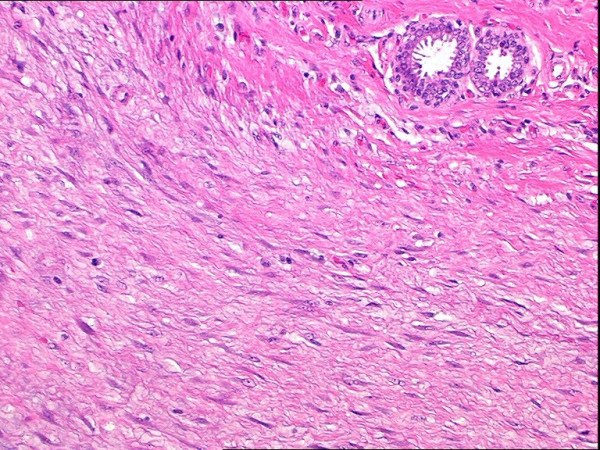
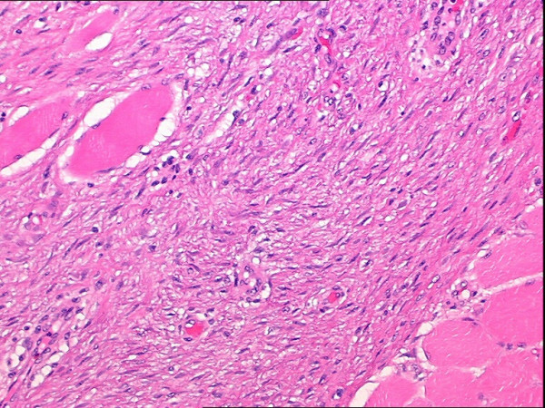
Background: Fibromatosis or desmoid tumor is a rare soft tissue tumor that lacks a metastatic potential, but is characterized by a locally aggressive and infiltrating growth pattern and a high propensity toward local recurrence if incompletely excised.
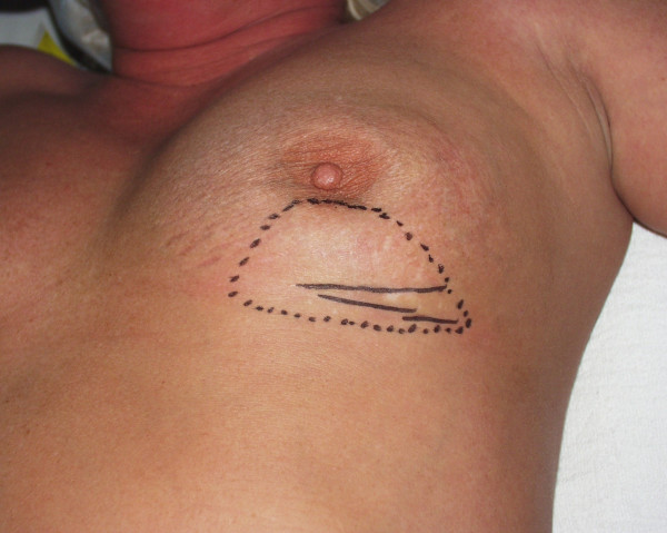
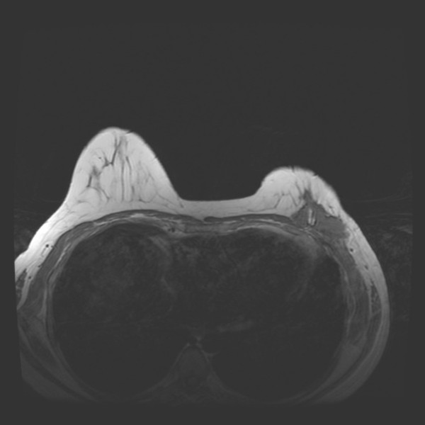
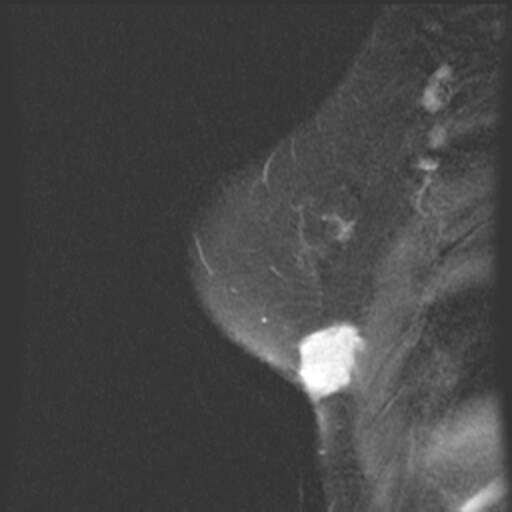
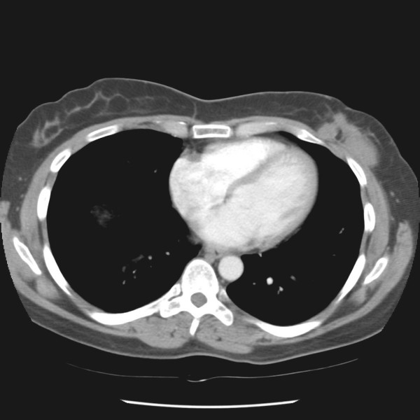
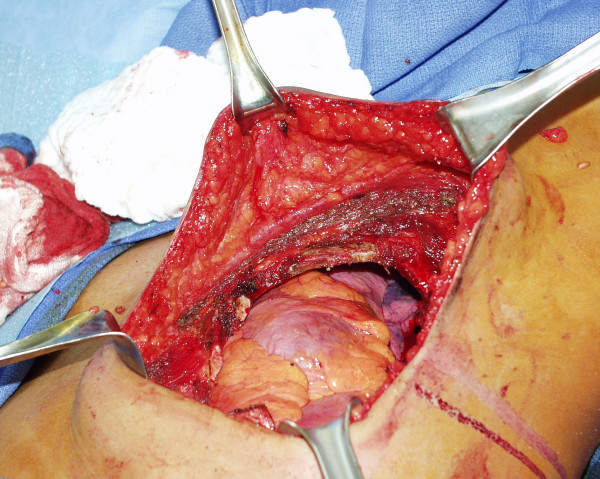
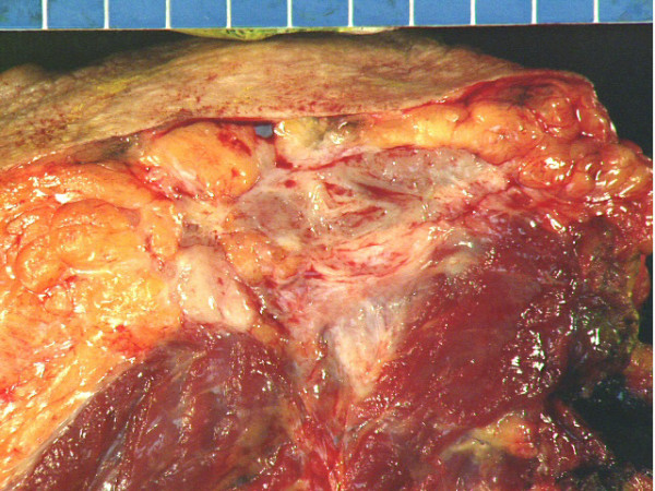
Case presentation: We report a patient with three post-surgical recurrences of fibromatosis of the breast over a seven year period. The fibromatosis was found to be involving the chest wall musculature and causing persistent and worsening pain. An aggressive operative strategy was undertaken, consisting of mastectomy with en bloc resection of the underlying chest wall musculature, ribs, and parietal pleura.
Conclusion: Aggressive surgical management of fibromatosis of the breast with suspected chest wall involvement is appropriate to attempt to obtain a long-term durable cure.
Figures
References
-
- Allen PW. The fibromatoses: a clinicopathologic classification based on 140 cases. Part I. Am J Surg Pathol. 1977;1:255–270. - PubMed
-
- Allen PW. The fibromatoses: a clinicopathologic classification based on 140 cases. Part II. Am J Surg Pathol. 1977;1:305–321. - PubMed
-
- Enzinger FM, Weiss SW. Fibromatosis. In: Enzinger FM, Weiss SW, editor. Soft Tissue Tumors. 4. St. Louis, MO, Mosby; 2001. pp. 320–329.
-
- Wargotz ES, Norris HJ, Austin RM, Enzinger FM. Fibromatosis of the breast. A clinical and pathological study of 28 cases. Am J Surg Pathol. 1987;11:38–45. - PubMed
-
- Kalisher L, Long JA, Peyster RG. Extra-abdominal desmoid of the axillary tail mimicking breast carcinoma. Am J Roentgenol. 1976;126:903–906. - PubMed
LinkOut - more resources
Full Text Sources
Research Materials
