. 2006 Jun 19;173(6):927-36.
doi: 10.1083/jcb.200512024. Epub 2006 Jun 12.

The Sec1p/Munc18 protein Vps45p binds its cognate SNARE proteins via two distinct modes

Affiliations

The Sec1p/Munc18 protein Vps45p binds its cognate SNARE proteins via two distinct modes

Lindsay N Carpp et al. J Cell Biol. .

Abstract

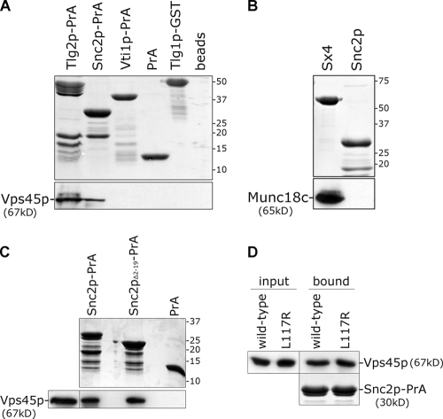
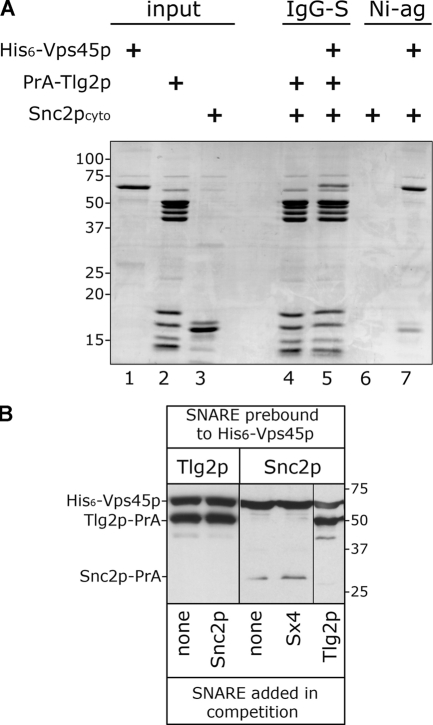
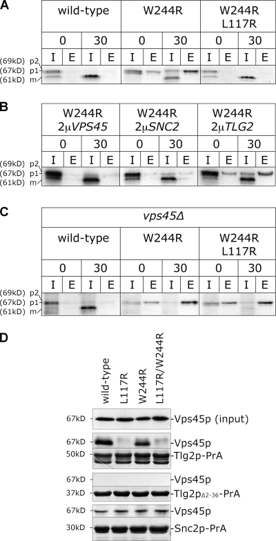
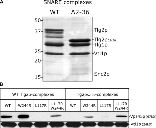
Sec1p/Munc18 (SM) proteins are essential for SNARE-mediated membrane trafficking. The formulation of unifying hypotheses for the function of the SM protein family has been hampered by the observation that two of its members bind their cognate syntaxins (Sxs) in strikingly different ways. The SM protein Vps45p binds its Sx Tlg2p in a manner analogous to that captured by the Sly1p-Sed5p crystal structure, whereby the NH2-terminal peptide of the Sx inserts into a hydrophobic pocket on the outer face of domain I of the SM protein. In this study, we report that although this mode of interaction is critical for the binding of Vps45p to Tlg2p, the SM protein also binds Tlg2p-containing SNARE complexes via a second mode that involves neither the NH2 terminus of Tlg2p nor the region of Vps45p that facilitates this interaction. Our findings point to the possibility that SM proteins interact with their cognate SNARE proteins through distinct mechanisms at different stages in the SNARE assembly/disassembly cycle.


Figures

Figure 1.
Figure 1.
Abrogation of Vps45p binding to Tlg2p does not perturb membrane traffic. (A) Sly1p and Vps45p sequences from S. cerevisiae were aligned using Clustalw software. A portion of the alignment containing the hydrophobic pocket of Sly1p into which the NH2-terminal peptide of Sed5p inserts is shown (residues 128–165 of Sly1p and residues 108–141 of Vps45p). The five residues that form the hydrophobic pocket of Sly1p are shaded. Asterisks indicate residues that are identical between the two proteins. Double dots indicate a strong similarity between amino acid residues, and a single dot indicates similarity. (B) Tlg2p-PrA immobilized on IgG-Sepharose was incubated with cell lysates prepared from vps45Δ cells (NOzY1) harboring plasmids encoding either HA-Vps45p (pCOG070) or HA-Vps45pL117R (pCOG071). Bound material was subject to immunoblot analysis using HA antibodies (10% of the lysate used was included; input). (C) Vps45p and bound proteins were immunoprecipitated from vps45Δ cells (NozY1) harboring plasmids encoding either HA-Vps45p (pCOG070) or HA-Vps45pL117R (pCOG071). The amount of Vps45p and Tlg2p present in the immunoprecipitates was assessed by immunoblotting (a sample of the lysate was also included in this analysis; 10% on the Vps45p blot and 0.5% on the Tlg2p blot). (D) CPY sorting was followed in wild-type (RPY10), vps45Δ (NozY2), and vps45Δ cells carrying a centromeric plasmid expressing either wild-type VPS45 (pNB706) or a version of the gene encoding Vps45pL117R (pNB708). Cells were pulse labeled for 5 min with [35S]methionine/cysteine. CPY was immunoprecipitated from both intracellular (I) and extracellular (E) fractions at the beginning (0) and end (30) of a 30-min chase period.
Figure 2.
Figure 2.
Vps45p binds directly to the v-SNARE Snc2p. Recombinant proteins purified from E. coli were immobilized on glutathione-agarose or IgG-Sepharose before incubation with either His6-tagged Vps45p or Munc18c purified from E. coli. After extensive washing, bound proteins were subject to SDS-PAGE followed by Coomassie blue staining (top) or immunoblotting with Vps45p or Munc18c antibodies. (A) Proteins comprising the cytosolic domains of Tlg2p, Snc2p, Vti1p, and Tlg1p fused to either PrA or GST were tested for their ability to bind His6-Vps45p. (B) The binding of Munc18c to Snc2p-PrA and GST-tagged syntaxin 4 (Sx4) was assessed. (C) The ability of Snc2p-PrA, Snc2pΔ2–19PrA, and the PrA moiety alone to bind His6-Vps45p was analyzed. (D) The ability of Snc2p-PrA to bind either His6-tagged Vps45p (wild type) or the same harboring the L117R mutation purified from bacteria was assessed and run next to 5% of the lysate used (input).
Figure 3.
Figure 3.
Tlg2p and Snc2p do not bind simultaneously to Vps45p. (A) ∼1 nmol Tlg2p-PrA immobilized on IgG-Sepharose was incubated with Snc2cyto alone (lane 4) or in addition to purified His6-Vps45p (lane 5). Protein mixtures were incubated at 4°C for ∼16 h, after which time proteins were eluted from IgG-Sepharose (IgG-S) after extensive washing with PBS. Eluted proteins were resolved by 12% SDS-PAGE and stained with Coomassie blue. Snc2cyto was incubated under the same conditions either with or without purified His6-Vps45p (lanes 6 and 7). An excess of Ni2+-agarose resin (Ni-ag) was added for the last hour of these incubations, after which time proteins that bound to the resin were detected by Coomassie blue staining after SDS-PAGE. Proportional samples of the input proteins were included on the same gel (lanes 1–3). Note that both Snc2cyto and His6-Vps45p were added in excess to Tlg2p-PrA (approximately five- and twofold, respectively). (B) ∼1 nmol His6-Vps45p was immobilized on Ni2+-agarose resin and incubated with either ∼1 nmol Tlg2p-PrA or Snc2p-PrA at 4°C for ∼16 h to prebind the SNARE. After extensive washing in PBS, either 50 nmol Snc2p-PrA, Tlg2p-PrA, or GST-Sx4 or no SNARE was added in a total volume of 1 ml. After incubation at 4°C for a further 16 h, material bound to the Ni2+-agarose resin was assessed by immunoblotting.
Figure 4.
Figure 4.
Tlg2p binding is required for the efficacy of a dominant-negative version of Vps45p. (A) CPY sorting was followed (as in Fig. 1) in wild-type cells (RPY10) harboring the following plasmids: wild-type (HA-Vps45p; pCOG070), W244R (HA-Vps45pW244; pCOG072), and L117R/W244R (HA-Vps45pL117R/W244R; pCOG073). (B) As in A, but with wild-type cells (RPY10) producing HA-Vps45pW244 from pCOG065 and also harboring the following plasmids: 2μVPS45 (pCOG070), 2μSNC2 (pCOG054), and 2μTLG2 (pHA-TLG2). (C) vps45Δ cells (NOzY2) producing HA-Vps45p and mutants thereof from the same plasmids as in A. (D) The binding of HA-Vps45p and indicated mutants from yeast cell lysates prepared from vps45Δ (NOzY1) cells transformed with pCOG070 (wild type), pCOG072 (W244R), pCOG071 (L117R), or pCOG073 (L117R/W244R) to Tlg2p-PrA, Tlg2pΔ2–36-PrA, and Snc2p-PrA was assessed as described in Fig. 1. Gels were also stained with Coomassie blue. The amount of Vps45p in 5% of each lysate used is also shown (input). I, intracellular; E, extracellular.
Figure 5.
Figure 5.
The NH2-terminal peptide of Tlg2p is not required for Vps45p to bind assembled SNARE complexes. (A) SNARE complexes containing either the entire cytosolic domain of Tlg2p (WT) or lacking residues 2–36 (Δ2–36) bound to Ni2+-agarose were resolved by SDS-PAGE on a 14% gel and were visualized by Coomassie staining. Note that the relatively weak band for Snc2p is caused by its poor Coomassie staining (McNew et al., 2000). The bands migrating between Tlg2p and Tlg1p in the wild-type lane are Tlg2p degradation products (confirmed by immunoblotting; not depicted). (B) The binding of HA-Vps45p and indicated mutants from yeast cell lysates described in Fig. 4 to the complexes represented in A was assessed as described in Materials and methods. Bound material was analyzed through immunoblotting with anti-HA antibodies and anti-Vti1p (to ensure that binding to equivalent amounts of complex was being assessed).
Figure 6.
Figure 6.
Vps45p associates with membranes through a manner distinct from the binding represented by the Sly1p–Sed5p crystal structure. vps45Δ (NOzY1) cells expressing HA-tagged versions of wild-type Vps45p (pNB706), W244R (pNB707), L117R (pNB708), or L117R/W244R (pNB709) from centromeric plasmids were fractionated into membrane pellet (P) and cytosol (S) fractions (W, whole cell extract). The amount of Vps45p in each of these fractions was analyzed through immunoblotting. Phosphoglycerate kinase (PGK) and AP were included in this analysis as markers for membranes and cytosol, respectively.

References

    1. Bracher, A., and W. Weissenhorn. 2002. Structural basis for the Golgi membrane recruitment of Sly1p by Sed5p. EMBO J. 21:6114–6124. - PMC - PubMed
    1. Bracher, A., A. Perrakis, T. Dresbach, H. Betz, and W. Weissenhorn. 2000. The X-ray crystal structure of neuronal Sec1 from squid sheds new light on the role of this protein in exocytosis. Structure. 8:685–894. - PubMed
    1. Bryant, N.J., and D.E. James. 2001. Vps45p stabilizes the syntaxin homologue Tlg2p and positively regulates SNARE complex formation. EMBO J. 20:3380–3388. - PMC - PubMed
    1. Bryant, N.J., and D.E. James. 2003. The Sec1p/Munc18 (SM) protein, Vps45p, cycles on and off membranes during vesicle transport. J. Cell Biol. 161:691–696. - PMC - PubMed
    1. Carr, C.M., E. Grote, M. Munson, F.M. Hughson, and P.J. Novick. 1999. Sec1p binds to SNARE complexes and concentrates at sites of secretion. J. Cell Biol. 146:333–344. - PMC - PubMed

Publication types

MeSH terms