. 2006 Jun 20;103(25):9542-7.
doi: 10.1073/pnas.0602250103. Epub 2006 Jun 12.

IRBIT, an inositol 1,4,5-trisphosphate receptor-binding protein, specifically binds to and activates pancreas-type Na+/HCO3- cotransporter 1 (pNBC1)

Affiliations

IRBIT, an inositol 1,4,5-trisphosphate receptor-binding protein, specifically binds to and activates pancreas-type Na+/HCO3- cotransporter 1 (pNBC1)

Kyoko Shirakabe et al. Proc Natl Acad Sci U S A. .

Abstract

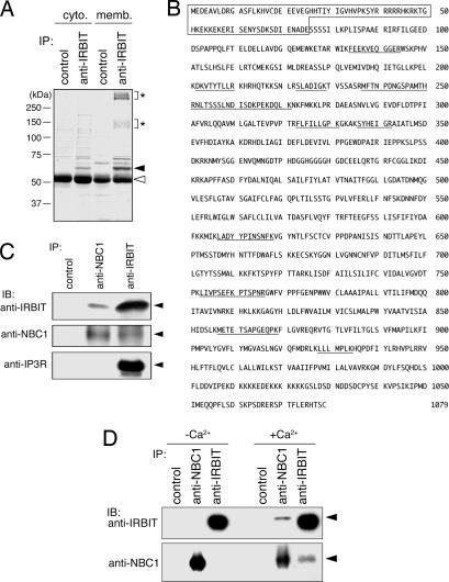
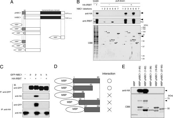
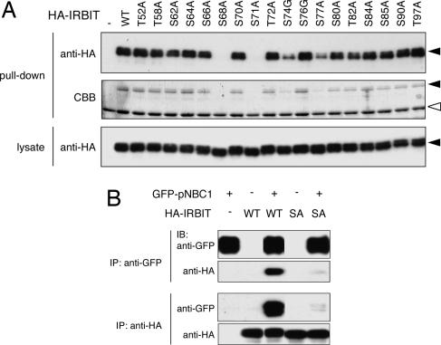
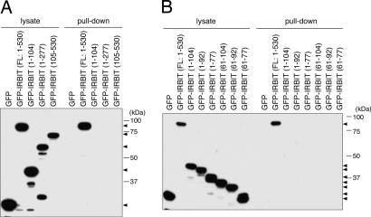
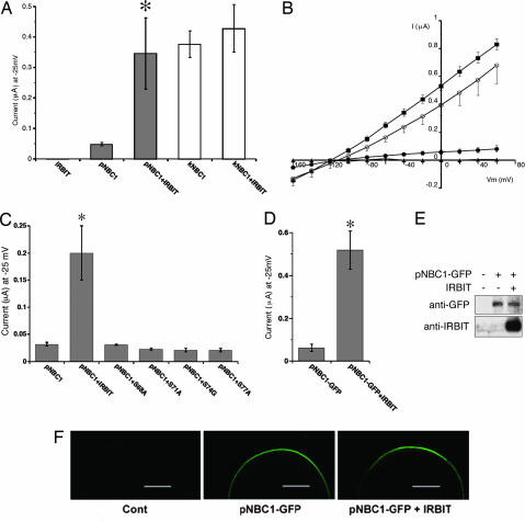
Inositol 1,4,5-trisphosphate (IP(3)) receptors (IP(3)Rs) are IP(3)-gated Ca(2+) channels that are located on intracellular Ca(2+) stores. We previously identified an IP(3)R binding protein, termed IP(3)R binding protein released with IP(3) (IRBIT). Because IRBIT is released from IP(3)R by physiological concentrations of IP(3), we hypothesized that IRBIT is a signaling molecule that is released from IP(3)R and regulates downstream target molecules in response to the production of IP(3). Therefore, in this study, we attempted to identify the target molecules of IRBIT, and we succeeded in identifying Na(+)/HCO(3)(-) cotransporter 1 (NBC1) as an IRBIT binding protein. Of the two major splicing variants of NBC1, pancreas-type NBC1 (pNBC1) and kidney-type NBC1 (kNBC1), IRBIT was found to bind specifically to pNBC1 and not to bind to kNBC1. IRBIT binds to the N-terminal pNBC1-specific domain, and its binding depends on the phosphorylation of multiple serine residues of IRBIT. Also, an electrophysiological analysis in Xenopus oocytes revealed that pNBC1 requires coexpression of IRBIT to manifest substantial activity comparable with that of kNBC1, which displays substantial activity independently of IRBIT. These results strongly suggest that pNBC1 is the target molecule of IRBIT and that IRBIT has an important role in pH regulation through pNBC1. Also, our findings raise the possibility that the regulation through IRBIT enables NBC1 variants to have different physiological roles.


Conflict of interest statement

Conflict of interest statement: No conflicts declared.

Figures

Fig. 1.
Fig. 1.
Identification of NBC1 as an IRBIT binding protein. (A) Mouse cerebella were collected and fractionated into cytoplasmic (cyto.) and membrane (memb.) fractions, and each fraction was subjected to immunoprecipitation (IP) with control rabbit IgG and rabbit anti-IRBIT Ab. Immunoprecipitates were subjected to SDS/PAGE using a 10% acrylamide gel, and the gel was stained with Coomassie brilliant blue (CBB). Filled and open triangles indicate the migration of endogenous IRBIT and IgG proteins, respectively. Asterisks indicate the migration of membrane fraction-specific IRBIT binding proteins. (B) Amino acid sequence of pNBC1. The peptides obtained by digestion of membrane fraction-specific IRBIT binding proteins (asterisks in A) are underlined. The N-terminal pNBC1-specific domain is boxed. (C) Mouse cerebellar membrane fraction was subjected to immunoprecipitation with rabbit control IgG, rabbit anti-NBC1, and rabbit anti-IRBIT Ab. Immunoprecipitates were subjected to Western blotting (IB) with guinea pig anti-IRBIT, rabbit anti-NBC1, and rat anti-IP3R Ab. Filled triangles indicate the migration of endogenous IRBIT, NBC1, and IP3R proteins. (D) COS-7 cells were lysed by lysis buffer with (+Ca2+) or without (−Ca2+) 2 mM CaCl2. Each lysate was subjected to immunoprecipitation with rabbit control IgG, rabbit anti-NBC1, and rabbit anti-IRBIT Ab, and immunoprecipitates were subjected to Western blotting with guinea pig anti-IRBIT and guinea pig anti-NBC1 Ab. Filled triangles indicate the migration of endogenous IRBIT and NBC1 proteins.
Fig. 2.
Fig. 2.
IRBIT specifically interacts with N-terminal pNBC1-specific domain. (A) Diagram of pNBC1, kNBC1, and NBC1 deletion mutants. MBP, maltose-binding protein. (B) COS-7 cells were transfected with empty vector (−) or hemagglutinin (HA)-tagged IRBIT (HA-IRBIT)-expressing plasmid (+). Cell lysates were incubated with each NBC1 deletion mutant protein, and bound proteins (pull-down) were subjected to Western blotting with anti-HA (Top) and rabbit anti-IRBIT Ab (Middle), and CBB staining (Bottom). Filled and open triangles indicate migrations of HA-IRBIT and endogenous IRBIT proteins, respectively. Filled circles indicate migrations of NBC1 deletion mutant proteins. (C) COS-7 cells were transfected with GFP-NBC1- and/or HA-IRBIT-expressing plasmid, cell lysates were subjected to immunoprecipitation (IP) with anti-GFP and anti-HA Ab, and immunoprecipitates were subjected to Western blotting (IB) with anti-GFP and anti-HA Ab. (D) Diagram of deletion mutants of pNBC1-specific domain. (E) HA-IRBIT-expressing COS-7 cell lysate was incubated with the indicated deletion mutant proteins of pNBC1-specific domain. Bound proteins were pulled down and subjected to Western blotting with anti-HA Ab (Upper) and CBB staining (Lower). Filled triangles indicate the migrations of HA-IRBIT proteins. Filled circles indicate migrations of deletion mutant proteins of pNBC1-specific domain.
Fig. 3.
Fig. 3.
Phosphorylation of IRBIT is required for the interaction with pNBC1. (A) COS-7 cells were transfected with expression plasmids encoding indicated serine/threonine-substituted mutants of IRBIT. Cell lysates were incubated with recombinant protein of pNBC1-specific domain (no. 5 in Fig. 2A), and bound proteins were pulled down and subjected to Western blotting with anti-HA Ab (Top) and CBB staining (Middle). Cell lysates were subjected to Western blotting with anti-HA Ab (Bottom). Filled and open triangles indicate the migrations of HA-IRBIT proteins and recombinant protein of pNBC1-specific domain, respectively. (B) COS-7 cells were transfected with indicated plasmids, and cell lysates were subjected to immunoprecipitation (IP) with indicated Abs, and immunoprecipitates were subjected to Western blotting (IB) with indicated Abs.
Fig. 4.
Fig. 4.
Conformation of IRBIT required for the interaction with pNBC1. (A and B) COS-7 cells were transfected with expression plasmids encoding GFP-fused IRBIT deletion mutants. Cell lysates were incubated with recombinant protein of pNBC1-specific domain (no. 5 in Fig. 2A), and bound proteins were pulled down and subjected to Western blotting with anti-GFP Ab. Filled triangles indicate the migrations of GFP-fused IRBIT deletion mutant proteins.
Fig. 5.
Fig. 5.
pNBC1 requires coexpression of IRBIT to manifest substantial activity comparable with that of kNBC1. (A) NBC1-mediated currents in Xenopus oocyte. Influxes of anion charges, induced by solution change from ND-96 to HCO3-containing solution, were measured at a holding potential of −25 mV. Numbers of observation were 5 (IRBIT), 9 (pNBC1), 11 (pNBC1 + IRBIT), 5 (kNBC1), and 5 (kNBC1 + IRBIT). ∗, P < 0.01 versus pNBC1. (B) Current–voltage (IV) relationship of NBC1 currents in oocytes injected with cRNA of IRBIT (filled triangles), pNBC1 (filled circles), pNBC1 + IRBIT (open circles), and kNBC1 (filled squires). Step pulses between Vm = −160 and 60 mV were applied in the absence and presence of an NBC1 inhibitor, 4,4′-diisothiocyanatostilbene-2,2′-disulfonic acid (0.4 mM). Reversal potentials were 108 ± 6 mV (pNBC1), 115 ± 13 mV (pNBC1 + IRBIT), and 117 ± 3 mV (kNBC1). (C) Effects of WT IRBIT (IRBIT) or IRBIT mutants on pNBC1 currents measured at a holding potential of −25 mV. Numbers of observation were 5 for each construct. ∗, P < 0.05 versus pNBC1. (D) Effect of IRBIT on the currents mediated by C-terminal GFP-tagged pNBC1 (pNBC1-GFP). Numbers of observation were 4 for each construct. ∗, P < 0.05 versus pNBC1-GFP. (E) Xenopus oocytes injected with indicated cRNAs were extracted, and extracts were subjected to Western blotting with anti-GFP and guinea pig anti-IRBIT Ab. (F) Surface expression of pNBC1-GFP observed by a confocal laser scanning microscopy. (Scale bars, 300 μm.)

References

    1. Berridge M. J. Nature. 1993;361:315–325. - PubMed
    1. Ando H., Mizutani A., Matsu-ura T., Mikoshiba K. J. Biol. Chem. 2003;278:10602–10612. - PubMed
    1. Soleimani M., Burnham C. E. J. Membr. Biol. 2001;183:71–84. - PubMed
    1. Igarashi T., Inatomi J., Sekine T., Cha S. H., Kanai Y., Kunimi M., Tsukamoto K., Satoh H., Shimadzu M., Tozawa F., et al. Nat. Genet. 1999;23:264–266. - PubMed
    1. Igarashi T., Inatomi J., Sekine T., Seki G., Shimadzu M., Tozawa F., Takeshima Y., Takumi T., Takahashi T., Yoshikawa N., et al. J. Am. Soc. Nephrol. 2001;12:713–718. - PubMed

Publication types