. 2006 Jun 20;103(25):9631-6.
doi: 10.1073/pnas.0600225103. Epub 2006 Jun 12.

The mutant leucine-zipper domain impairs both dimerization and suppressive function of Foxp3 in T cells

Affiliations

The mutant leucine-zipper domain impairs both dimerization and suppressive function of Foxp3 in T cells

Wook-Jin Chae et al. Proc Natl Acad Sci U S A. .

Abstract

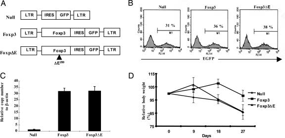
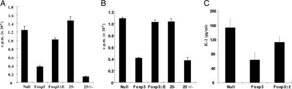
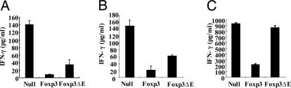
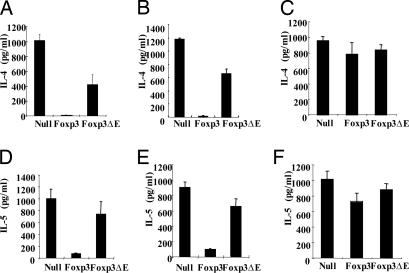
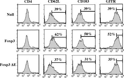
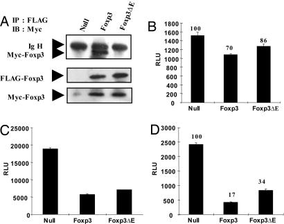
Regulatory T cells that express the Foxp3 transcription factor play important roles in preventing autoimmune diseases. Although several studies have demonstrated that the lack of the forkhead DNA-binding domain of Foxp3 caused severe autoimmune disease in scurfy mutant mice, the other functional domains of Foxp3 are less well characterized. Here, we show that the deletion of glutamic acid (DeltaE250) in the leucine-zipper domain of Foxp3 causes a loss of hyporesponsiveness when compared with wild-type Foxp3 upon antigenic stimulation. CD4 T cells that ectopically express the glutamic acid mutant show significant losses of suppressor activity both in vitro and in vivo. We also demonstrate that regulation of both Th1- and Th2-type cytokine secretion in CD4 T cells that express wild-type Foxp3 is significantly altered by the deletion of glutamic acid. Defects are also observed in the expression of adhesion molecules, such as l-selectin (CD62L) and CD103, suggesting an important role of glutamic acid in the migratory behavior of regulatory T cells. Finally, this mutation reduces transcriptional repressor activity and impairs the homodimerization of Foxp3. Taken together, our results provide insight into the mechanism that controls autoimmune diseases via the deletion of this single glutamic acid residue in the leucine-zipper domain of Foxp3.


Conflict of interest statement

Conflict of interest statement: No conflicts declared.

Figures

Fig. 1.
Fig. 1.
The ΔE mutation in the leucine-zipper domain of Foxp3 prevents generation of functional regulatory T cells. (A) Each Foxp3 and Foxp3 ΔE cDNA was cloned into MSCV 2.2 retroviral vectors containing the marker GFP. (B) CD4+CD25− T cells from C57BL/6 mice were transduced with retroviruses, and GFP-positive cells were then sorted. (C) GFP-positive cells were analyzed by real-time PCR for Foxp3 expression. (D) The effect of transduced CD4+ T cells on the progression of IBD was monitored by mean body weight. Closed circles, null vector; closed squares, wild-type Foxp3; closed triangles, Foxp3ΔE.
Fig. 2.
Fig. 2.
Effects of ΔE mutation on proliferation, suppressor function, and IL2 secretion. The ΔE mutation in Foxp3 leads to loss of suppressor function of Foxp3-transduced CD4 cells cultured alone (A) or with purified CD4+CD25− T cells (B). (C) IL-2 secretion was measured in transduced CD4 cells that were cultured alone.
Fig. 3.
Fig. 3.
Effects of ΔE mutation on IFN-γ (Th1 cytokine) secretion. The ΔE mutation in Foxp3 impairs Foxp3-induced inhibition of CD4 T cells cultured alone (A) or with either purified CD4+CD25− T cells (B) or Th1 differentiated CD4 T cells (C).
Fig. 4.
Fig. 4.
Effects of ΔE mutation on Th2 cytokines. The ΔE mutation in Foxp3 attenuates Foxp3-induced suppression of IL-4 and IL-5 secretion in CD4 cells (A and D) cultured alone or with CD4+CD25− T cells (B and E). Cytokine expression by coculture of transduced CD4 cells with Th1- and Th2-differentiated T cells (C and F).
Fig. 5.
Fig. 5.
Deletion of a functional glutamic acid residue down-regulates cell surface markers including adhesion molecule. Each group of GFP-positive cells was sorted and stained with APC-conjugated anti-CD4, phycoerythrin-conjugated anti-CD62L, anti-CD103, and anti-GITR Abs. A representative result from three independent experiments is shown.
Fig. 6.
Fig. 6.
The glutamic acid residue in the leucine-zipper domain of Foxp3 is required for homodimerization. (A) FLAG- and Myc-tagged Foxp3 or Foxp3ΔE were cotransfected for 48 h. After harvesting of cells, lysates were incubated with FLAG antibody (M2)-conjugated agarose beads, washed and boiled with sample buffer for SDS/PAGE. Immunoprecipitates were analyzed by anti-Myc Ab. Lysates were also analyzed for protein expression by using anti-FLAG and anti-Myc Abs. RLU, relative luciferase unit. (B–D) Each null vector, Foxp3, and Foxp3 ΔE was cotransfected with the FKH luciferase construct in Jurkat T cells with lipid-mediated transfection (B). After 48 h, luciferase activity was measured (C and D). Each NFAT and NF-κB luciferase construct was cotransfected with null vector, Foxp3, or Foxp3ΔE in HEK293 cells. After 48-h transfection, cells were harvested and assessed for luciferase activity. All samples were normalized by cotransfection and analysis of TK-Renilla activity. A representative result from at least three independent experiments is shown.

Similar articles

Cited by

References

    1. Sakaguchi S. Annu. Rev. Immunol. 2004;22:531–562. - PubMed
    1. Gavin M., Rudensky A. Curr. Opin. Immunol. 2003;15:690–696. - PubMed
    1. Baecher-Allan C., Viglietta V., Hafler D. A. Semin. Immunol. 2004;16:89–98. - PubMed
    1. Schubert L. A., Jeffery E., Zhang Y., Ramsdell F., Ziegler S. F. J. Biol. Chem. 2001;276:37672–37679. - PubMed
    1. Hori S., Nomura T., Sakaguchi S. Science. 2003;299:1057–1061. - PubMed

Publication types

MeSH terms