. 2006 Jun;20(8):1131-41.
doi: 10.1096/fj.05-5483com.

Overexpression of SUR2A generates a cardiac phenotype resistant to ischemia

Affiliations

Overexpression of SUR2A generates a cardiac phenotype resistant to ischemia

Qingyou Du et al. FASEB J. 2006 Jun.

Abstract

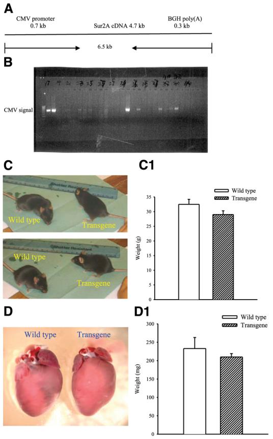
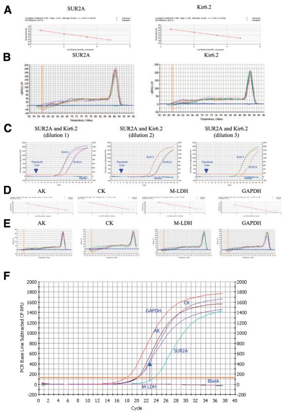
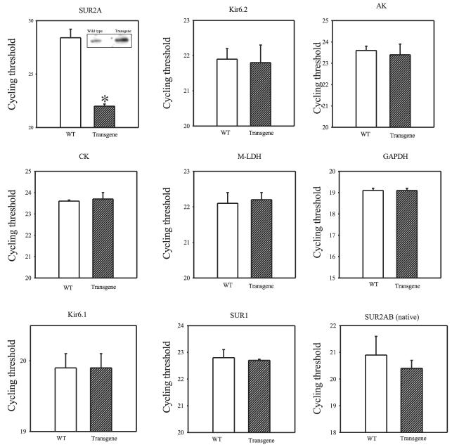
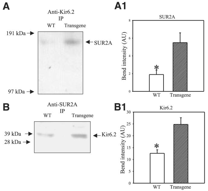
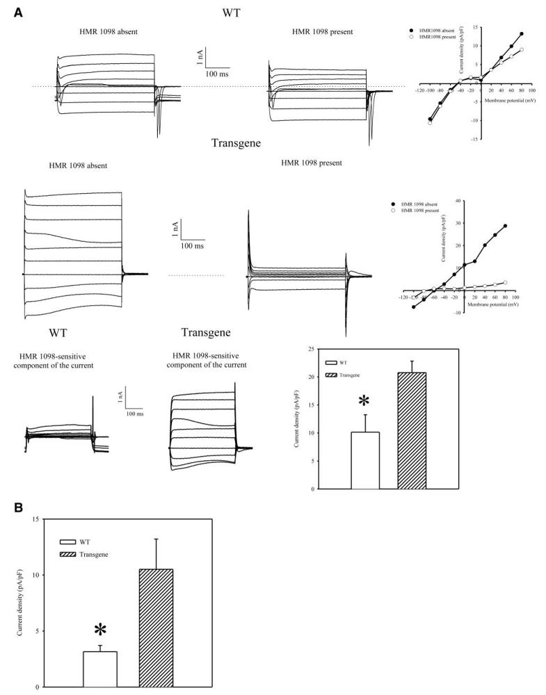
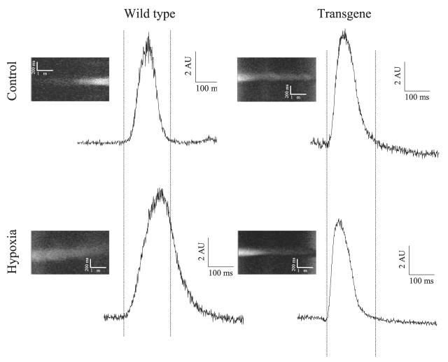
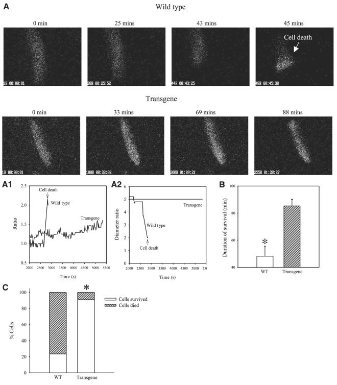
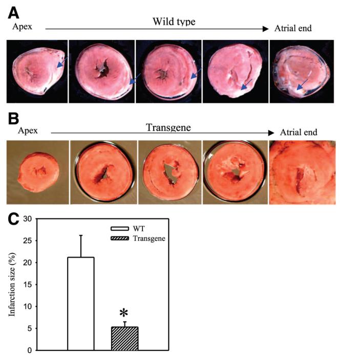
ATP-sensitive K+ (K(ATP)) channels are present in the sarcolemma of cardiac myocytes where they link membrane excitability with the cellular bioenergetic state. These channels are in vivo composed of Kir6.2, a pore-forming subunit, SUR2A, a regulatory subunit, and at least four accessory proteins. In the present study, real-time RT-PCR has demonstrated that of all six sarcolemmal K(ATP) channel-forming proteins, SUR2A was probably the least expressed protein. We have generated mice where the SUR2A was under the control of a cytomegalovirus promoter, a promoter that is more efficient than the native promoter. These mice had an increase in SUR2A mRNA/protein levels in the heart whereas levels of mRNAs of other channel-forming proteins were not affected at all. Imunoprecipitation/Western blot and patch clamp electrophysiology has shown an increase in K(ATP) channel numbers in the sarcolemma of transgenic mice. Cardiomyocytes from transgenic mice responded to hypoxia with shortening of action membrane potential and were significantly more resistant to this insult than cardiomyocytes from the wild-type. The size of myocardial infarction in response to ischemia-reperfusion was much smaller in hearts from transgenic mice compared to those in wild-type. We conclude that overexpression of SUR2A generates cardiac phenotype resistant to hypoxia/ischemia/reperfusion injury due at least in part to increase in levels of sarcolemmal K(ATP) channels.


Figures

Figure 1
Figure 1
Generation of transgenic mice. A) Schematic representation of the CMV-SUR2A construct used to generate transgenic mice. B) Founder mice were checked for successful integration of the transgene CMV-SUR2A by PCR. PCR products obtained with CMV-specific primers using mouse genomic DNA as template. C, D) Photographs of wild-type and transgenic mice (C) and hearts (D) from these mice. C1 and D1 correspond to panels C and D, respectively. Each bar represents mean ± se (n=6–16).
Figure 2
Figure 2
Levels of mRNA of sarcolemmal KATP channel subunits and accessory proteins in the mouse heart. A) Standard curve for the real-time PCR amplification of SUR2A and Kir6.2 cDNA. Shown is a linear regression plot of the cross point (cycle number) vs. the logarithm of cDNA amounts (40 pg, 200 pg, 1 ng, 5 ng, 25 ng). Each point represents an experiment. B) Melting curve analysis of Sur2A and Kir6.2 amplicons. Shown are plots of fluorescence (−dF1/dT) vs. temperature with different amounts of cDNA template (40 pg, 200 pg, 1 ng. 5 ng, 25 ng) done in duplicate. The presence of a single peak is consistent with the formation of a single amplicon and indicates the lack of primer-dimer formation. C) Representative progress curves done in duplicate for the real-time PCR amplification of SUR2A and Kir6.2 cDNA (dilution 1: 25 ng of template; dilution 2: 5 ng of template; dilution 3: 1 ng of template). D) Standard curve for the real-time PCR amplification of adenylate kinase type 1 (AK), creatine kinase type 1 (CK), muscle form of lactate dehydrogenase (m-LDH) and glyceraldehyde 3-phosphate dehydrogenase (GAPDH) cDNA. Shown is a linear regression plot of the cross point (cycle number) vs. the logarithm of cDNA amounts (80 pg, 400 pg, 2 ng, 10 ng, 50 ng). Each point represents an experiment. E) Melting curve analysis of AK, CK, m-LDH, and GAPDH amplicons. Shown are plots of fluorescence (−dF1/dT) vs. temperature with different amounts of cDNA template (80 pg, 400 pg, 2 ng, 10 ng, 50 ng) done in duplicate. F) Representative progress curves for the real-time PCR amplification of SUR2A, AK, CK, m-LDH, and GAPDH cDNA (5 ng of template was used).
Figure 3
Figure 3
Levels of mRNA of sarcolemmal KATP channel subunits and accessory proteins in transgenic and wild-type hearts. Bar graphs showing cycle threshold for the real-time PCR amplification of SUR2A, Kir6.2, AK, CK, m-LDH, GAPDH, Kir6.1, SUR1, and natively expressed SUR2A/SUR2B (SUR2AB, native) cDNA (25 ng of template was used) from wild-type (WT) and transgenic mice. Inset in Fig. 3 (SUR2A): Western blot of total protein extract with anti-SUR2 Ab. Each bar represents mean ± se (n=3–6). *P < 0.01.
Figure 4
Figure 4
A, B) Western blot with anti-Kir6.2 and anti-SUR2 Ab of anti-Kir6.2 (A) and anti-SUR2A (B) immunoprecipitate pellets (IP) from cardiac membrane fractions obtained from wild-type (WT) and transgenic mice. A1, B1). Graphs corresponding to panels A and B, respectively. Each bar represents mean ± se (n=3 for each). *P < 0.01.
Figure 5
Figure 5
Whole cell K+ currents in cells from wild-type (WT) and transgenic mice. A) Superimposed membrane currents evoked by identical families of 400 ms voltage pulses (from −100 mV to 80 mV; holding potential was −40 mV) in cells that were first maintained in the absence of HMR 1098, then exposed to 100 μM HMR 1098 for 2 min. Scatter-line graphs on the right correspond to the recordings on the left. Dotted lines correspond to the zero current concentration. B) Current densities at 80 mV in the absence of 100 μM HMR 1098 in cardiac cells from wild-type (WT) and transgenic mice. Each bar represents mean ± se (n=6–7). *P < 0.05.
Figure 6
Figure 6
The response of cardiac action membrane potential to hypoxia in wild-type and transgenic mice. Original line scans of di-8-ANEPPS-loaded cardiomyocytes and corresponding time courses under depicted conditions. AU = arbitrary units.
Figure 7
Figure 7
Resistance of cardiomyocytes from wild-type and transgenic mice to hypoxia. A) Epifluorescent images of Fura-2-loaded cardiomyocytes exposed to hypoxia. ×40. Time course of Fura-2 ratio (A1) and cell diameters (A2) corresponding to experiments in panel A. B) Average survival time of wild-type and transgenic cardiomyocytes exposed to hypoxia. Bars represent mean ± se (n=17–18). *P < 0.01. C) Percentage of cells from wild-type or transgenic mice that died/survived, n = 17–18. *P < 0.01. Note that cardiomyocytes from transgenic mice are more resistant to hypoxia then cardiomyocytes from wild-type mice.
Figure 8
Figure 8
Resistance of wild-type and transgenic hearts to ischemia-reperfusion. A, B) Typical photographs of myocardial slices from wild-type and transgenic mice exposed to ischemia-reperfusion. Infarcted areas are pale/gray (indicated also with the blue arrows) whereas viable myocardium is dark/red. C) Myocardial infarct size expressed as a percentage of area at risk zone (n=6–17). *P < 0.01. Note that hearts from transgenic mice are more resistant to myocardial infarction then hearts from wild-type mice.

References

    1. Kloner RA, Jennings RB. Consequences of brief ischaemia: stunning, preconditioning and their clinical implications: part 2. Circulation. 2001;104:3158–3167. - PubMed
    1. Kloner RA, Rezkalla SH. Cardiac protection during acute myocardial infarction: where do we stand in 2004? J. Am. Coll. Cardiol. 2004;44:276–286. - PubMed
    1. Noma A. ATP-regulated K+ channels in cardiac muscle. Nature. 1983;305:147–148. - PubMed
    1. Hanley PJ, Daut J. KATP channels and preconditioning: A re-examination of the role of mitochondrial KATP channels and an overview of alternative mechanisms. J. Mol. Cell. Cardiol. 2005;39:17–50. - PubMed
    1. Kane GC, Liu XK, Yamada S, Olson TM, Terzic A. Cardiac KATP channels in health and disease. J. Mol. Cell. Cardiol. 2005;38:937–943. - PMC - PubMed

Publication types

MeSH terms

LinkOut - more resources