Review
. 2006 May-Jun;8(5-6):847-67.
doi: 10.1089/ars.2006.8.847.

Posttranslational modifications in Cu,Zn-superoxide dismutase and mutations associated with amyotrophic lateral sclerosis

Affiliations
Review

Posttranslational modifications in Cu,Zn-superoxide dismutase and mutations associated with amyotrophic lateral sclerosis

Yoshiaki Furukawa et al. Antioxid Redox Signal. 2006 May-Jun.

Abstract

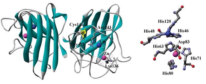
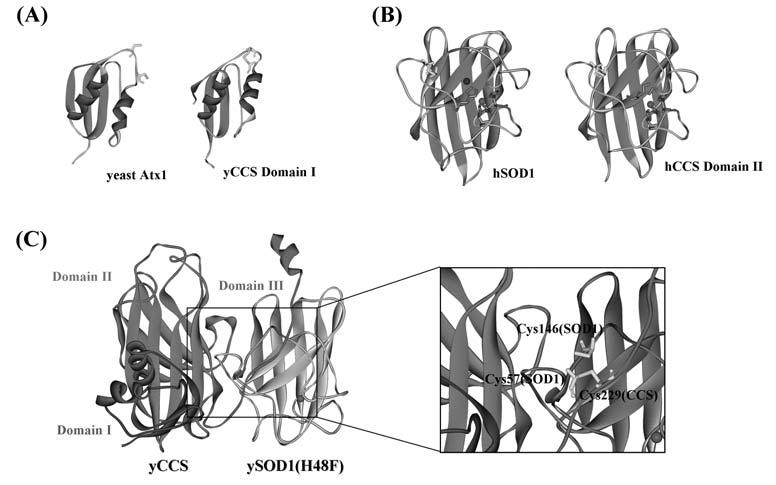
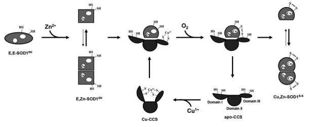
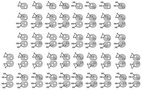
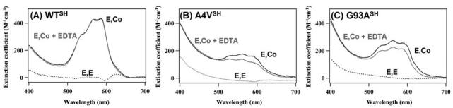
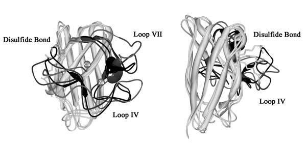
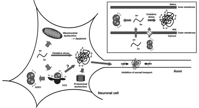
Activation of the enzyme Cu,Zn-superoxide dismutase (SOD1) involves several posttranslational modifications including copper and zinc binding, as well as formation of the intramolecular disulfide bond. The copper chaperone for SOD1, CCS, is responsible for intracellular copper loading in SOD1 under most physiological conditions. Recent in vitro and in vivo assays reveal that CCS not only delivers copper to SOD1 under stringent copper limitation, but it also facilitates the stepwise conversion of the disulfide-reduced immature SOD1 to the active disulfide-containing enzyme. The two new functions attributed to CCS, (i.e., O(2)-dependent sulfhydryl oxidase- and disulfide isomerase-like activities) indicate that this protein has attributes of the larger class of molecular chaperones. The CCS-dependent activation of SOD1 is dependent upon oxygen availability, suggesting that the cell only loads copper and activates this enzyme when O(2)-based oxidative stress is present. Thiol/disulfide status as well as metallation state of SOD1 significantly affects its structure and protein aggregation, which are relevant in pathologies of a neurodegenerative disease, amyotrophic lateral sclerosis (ALS). The authors review here a mechanism for posttranslational activation of SOD1 and discuss models for ALS in which the most immature forms of the SOD1 polypeptide exhibits propensity to form toxic aggregates.


Figures

FIG. 1
FIG. 1
Crystal structure of human SOD1 shown in a ribbon model (PDB ID: 1HL5). Cu, Zn ion and intramolecular disulfide bond are shown on ball and stick, respectively. Coordination structure around Cu and Zn ion is also shown in the right panel.
FIG. 2
FIG. 2
Crystal structures of CCS. (A) Domain I of CCS (PDB ID: 1QUP) has a homologous structure with another copper chaperone, Atx1 (PDB ID: 1FES). (B) Domain II of human CCS (PDB ID: 1DO5) is similar to human SOD1 (PDB ID: 1HL5). (C) Heterodimer structure between yeast SOD1 with the mutation, H48F, and yeast CCS (PDB ID: 1JK9). All three domains in CCS are identified in this crystal structure. Magnified image of the interfacial region containing intermolecular disulfide bond between SOD1 and CCS is also shown.
FIG. 3
FIG. 3
Our proposed model for the SOD1 activation cycle by Cu-CCS in yeast. Cu-CCS will interact with the Zn-bound and disulfide-reduced form of SOD1, E,Zn-SOD1SH. Yeast SOD1 favors monomeric state when the disulfide bond is reduced. Oxygen attacks the SOD1-CCS heterodimer to trigger the copper transfer, resulting in the formation of the intermolecular disulfide bond. Then the disulfide isomerization results in the intramolecular disulfide bond in SOD1 to form the dimeric holo-SOD1.
FIG. 4
FIG. 4
44 canonical microstates in SOD1. SOD1 microstates are dependent upon copper and zinc binding, intramolecular disulfide bond, and dimerization.
FIG. 5
FIG. 5
ALS-associated mutations in SOD1 cause the reduction of affinity for a Zn ion. Electronic absorption spectra of E,E-hSOD1SH (A: wild-type, B: A4V, C: G93A) in 50 mM HEPES, 1 mM tris-(2-carboxyethyl)phosphine, pH 7.4, with an equimolar CoCl2 at 20°C (solid black curve). 0.1 mM EDTA was further added to the E,Co-hSOD1SH samples, and the spectrum was obtained after 1 h incubation at 20°C (solid gray curve). Spectra of E,E-hSOD1SH alone were also shown as dotted curves. Molar extinction coefficient shown in the figure was calculated based upon the absorption at 280 nm, and the following protein concentration was used for the measurements: 67.3 μM wild-type, 74.5 μM A4V, 78.2 μM G93A hSOD1.
FIG. 6
FIG. 6
Superimposed images of E,E-, E,Znand Cu,Zn-hSOD1S-S structures (PDB ID: 1RK7, 1KMG, and 1BA9, respectively). An image from a different angle is also shown in the right panel. The conserved disulfide bond is shown in stick, and the Loops IV and VII are also highlighted by color; light gray, dark gray, and black in E,E-, E,Zn-, and Cu,Zn-hSOD1S-S, respectively.
FIG. 7
FIG. 7
Proposed processing of SOD1 protein in neuronal cells. Unmodified SOD1 polypeptide acquires a copper ion and a disulfide bond by forming a heterodimer with CCS, resulting in an enzymatic activation of SOD1. The free thiol groups on the SOD1 polypeptide can also be oxidized to form the disulfide-linked protein aggregates, which would lead to dysfunctions of proteasome and/or axonal transport. (Inset) The disulfide-reduced form of SOD1 can cross the outer membrane of mitochondria, but the disulfide form cannot. Due to the highly oxidative environment in the mitochondrial IMS, the imported SOD1 polypeptide is susceptible to disulfide crosslinks and aggregation in the absence of any posttranslational modifications.

References

    1. Abernethy JL, Steinman HM, Hill RL. Bovine erythrocyte superoxide dismutase. Subunit structure and sequence location of the intrasubunit disulfide bond. J Biol Chem. 1974;249:7339–7347. - PubMed
    1. Ahmad S, Gromiha M, Fawareh H, Sarai A. ASAView: database and tool for solvent accessibility representation in proteins. BMC Bioinformatics. 2004;5:51. - PMC - PubMed
    1. Andreassen OA, Dedeoglu A, Klivenyi P, Beal MF, Bush AI. N-acetyl-l-cysteine improves survival and preserves motor performance in an animal model of familial amyotrophic lateral sclerosis. Neuroreport. 2000;11:2491–2493. - PubMed
    1. Aoki M, Ogasawara M, Matsubara Y, Narisawa K, Nakamura S, Itoyama Y, Abe K. Mild ALS in Japan associated with novel SOD mutation. Nat Genet. 1993;5:323–324. - PubMed
    1. Arai K, Iizuka S, Tada Y, Oikawa K, Taniguchi N. Increase in the glucosylated form of erythrocyte Cu-Zn-superoxide dismutase in diabetes and close association of the nonenzymatic glucosylation with the enzyme activity. Biochim Biophys Acta. 1987;924:292–296. - PubMed