

Expression levels of Mycobacterium tuberculosis antigen-encoding genes versus production levels of antigen-specific T cells during stationary level lung infection in mice
- PMID: 16771854
- PMCID: PMC1782281
- DOI: 10.1111/j.1365-2567.2006.02355.x
Expression levels of Mycobacterium tuberculosis antigen-encoding genes versus production levels of antigen-specific T cells during stationary level lung infection in mice
Abstract
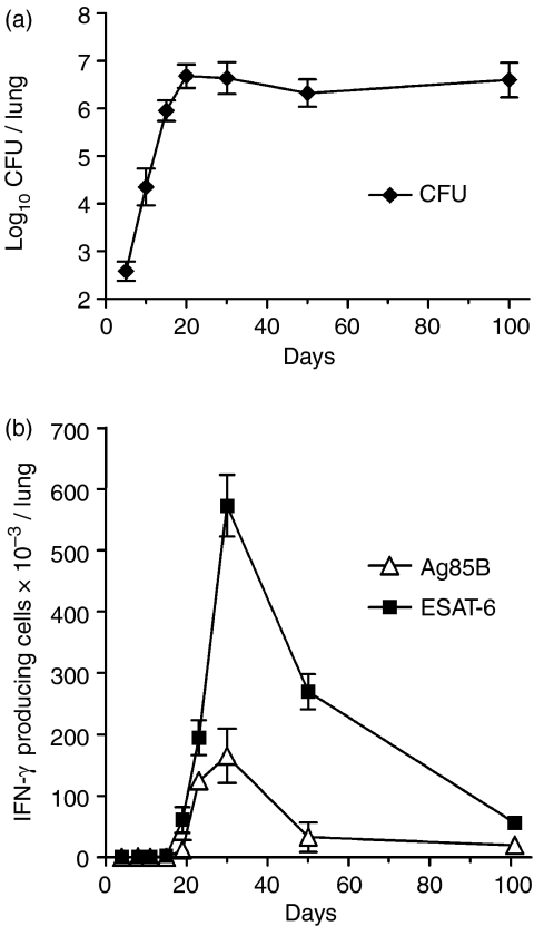
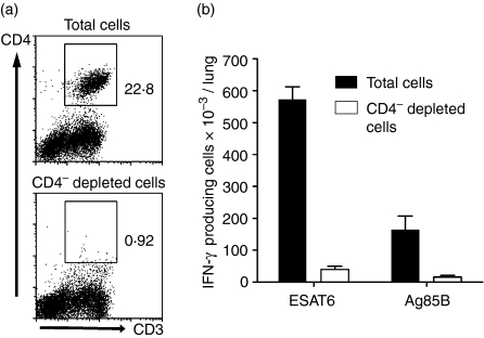
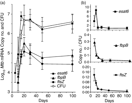
Mycobacterium tuberculosis lung infection in mice was controlled at an approximately stationary level after 20 days of log linear growth. Onset of stationary level infection was associated with the generation by the host of T helper type 1 (Th1) immunity, as evidenced by the accumulation of CD4 Th1 cells specific for the early secretory antigen (ESAT-6) of M. tuberculosis encoded by esat6, and for a mycolyl transferase (Ag85B) encoded by fbpB. CD4 T cells specific for these antigens were maintained at relatively high numbers throughout the course of infection. The number of CD4 T cells generated against ESAT-6 was larger than the number generated against Ag85B, and this was associated with a higher transcription level of esat6. The total number of transcripts of esat6 increased during the first 15 days of infection, after which it decreased and then approximately stabilized at 10(6.5) per lung. The total number of fbpB transcripts increased for 20 days of infection before decreasing and then approximately stabilizing at 10(4.8) per lung. The number of transcripts of esat6 per colony-forming unit of M. tuberculosis fell from 8.6 to 0.8 after day 15, and of fbpB from 0.3 to less than 0.02 after day 10, suggesting that at any given time during stationary level infection the latter gene was expressed by a very small percentage of bacilli. Expressed at an even lower level was an M. tuberculosis replication gene involved in septum formation (ftsZ), indicating that there was no significant turnover of the M. tuberculosis population during stationary level infection.
Figures
References
-
- Corbett EL, Watt CJ, Walker N, Maher D, Williams BG, Raviglione MC, Dye C. The growing burden of tuberculosis: global trends and interactions with the HIV epidemic. Arch Intern Med. 2003;163:1009–21. - PubMed
-
- Flynn JL, Chan J. Immunology of tuberculosis. Annu Rev Immunol. 2001;19:93–129. - PubMed
-
- North RJ, Jung YJ. Immunity to tuberculosis. Annu Rev Immunol. 2004;22:599–623. - PubMed
-
- Britton WJ, Palendira U. Improving vaccines against tuberculosis. Immunol Cell Biol. 2003;81:34–45. - PubMed
Publication types
MeSH terms
Substances
Grants and funding
LinkOut - more resources
Full Text Sources
Other Literature Sources
Research Materials
