. 2006 Jun;118(2):216-23.
doi: 10.1111/j.1365-2567.2006.02359.x.

Natural killer cells prime the responsiveness of autologous CD4+ T cells to CTLA4-Ig and interleukin-10 mediated inhibition in an allogeneic dendritic cell-mixed lymphocyte reaction

Affiliations

Natural killer cells prime the responsiveness of autologous CD4+ T cells to CTLA4-Ig and interleukin-10 mediated inhibition in an allogeneic dendritic cell-mixed lymphocyte reaction

Ashley Newland et al. Immunology. 2006 Jun.

Abstract

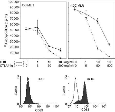
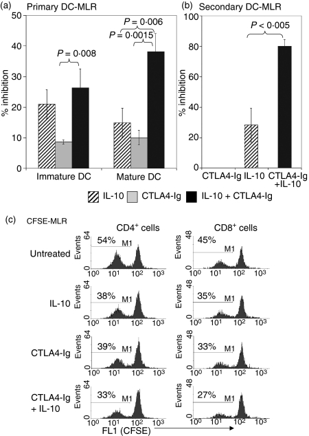
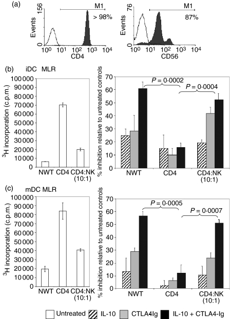
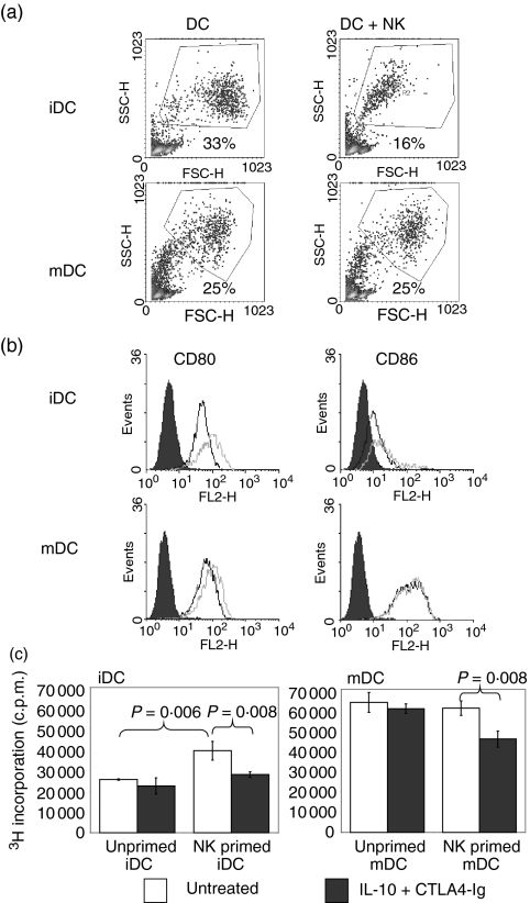
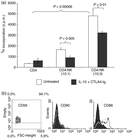
Cytotoxic T-lymphocyte antigen 4 immunoglobulin (CTLA4-Ig) and interleukin (IL)-10 are immunomodulatory molecules which target CD28 costimulation by acting either directly or indirectly on the CD80/86 receptors on dendritic cells (DCs). This study examined the effect of combined treatment with CTLA4-Ig and IL-10 on T-cell responsiveness in a dendritic cell-mixed lymphocyte reaction (DC-MLR). T cells derived from nylon wool enrichment (NWT cells) demonstrated 15% (P = 0.006) and 10% (P = 0.0015) inhibition of proliferation with suboptimal doses of IL-10 (5 ng/ml) and CTLA4-Ig (20 ng/ml), respectively. Combined treatment with both agents resulted in 38% inhibition (P = 0.004) of the MLR response compared with untreated controls. In contrast to NWT cells, which consisted of CD4+, CD8+ and CD56+ (NK) cells, purified CD4+ T cells were less responsive to immunomodulation by CTLA4-Ig and IL-10. Repletion of the CD4+ T cells with NK cells restored IL-10 and CTLA4-Ig mediated immunomodulation, suggesting a role for NK cells in the regulation of DC-T-cell interactions. The specific effect of NK cells on DC activation was demonstrated by CD80 up-regulation on DCs in the absence of T cells. However, in the absence of DCs, NK cells augmented the proliferation of autologous CD4+ T cells stimulated by anti-CD3 monoclonal antibody (mAb), which was blocked by CTLA4-Ig. It is proposed that, in the MLR, immunomodulation by suboptimal CTLA4-Ig and IL-10 is influenced by cellular interactions of NK cells with DCs and T cells involving DC lysis and costimulation. Thus, NK cells prime both DCs and T cells to low doses of CTLA4-Ig and IL-10 during alloimmune responses, providing evidence for the potential interaction between innate and adaptive immunity.


Figures

Figure 1
Figure 1
Dose–response effects of interleukin (IL)-10 and CTLA4-Ig in the DC-MLR. IL-10 (solid line) and CTLA4-Ig (dashed line) were titrated with 10-fold dilutions in the DC-MLR. Nylon wool T cells were used as the responder population at a stimulator:responder (S:R) ratio of 1 : 100. All samples were run in triplicate and proliferation was measured by [3H] thymidine incorporation, with results presented as counts per minute (c.p.m.) ± standard deviation (SD). The maturation status of the DCs was demonstrated by flow cytometric analysis by direct staining with anti-CD83-phycoerythrin (PE) (shaded) and with the negative control, isotype-matched PE-conjugated monoclonal antibody (unshaded). iDC, immature dendritic cell; mDC, mature dendritic cell.
Figure 2
Figure 2
Combined treatment of interleukin (IL)-10 and CTLA4-Ig inhibited the DC-MLR. Nylon wool T cells were used in the DC-MLR at a stimulator:responder (S:R) ratio of 1 : 100. (a) The percentage inhibition of IL-10 (5 ng/ml) and CTLA4 (20 ng/ml) alone or in combination in the immature or mature DC-MLR. (b) The percentage inhibition in a secondary MLR. T cells from the primary MLR were isolated and used for restimulation in a secondary MLR in the absence of both immunomodulatory agents. Proliferation in (a) and (b) was measured by [3H] thymidine incorporation, with results presented as the percentage inhibition compared with untreated controls. P-values were determined by unpaired Student's t-test. All samples were run in triplicate, and data shown for (a) and (b) are representative of 12 and three independent experiments, respectively. (c) Inhibition of CD4+ and CD8+ T-cell proliferation by IL-10 and CTLA4-Ig in a CFSE-MLR. Nylon wool T cells were stained with CFSE and added to DC stimulators. After 5 days in culture, cells were stained with anti-CD4-phycoerythrin (PE) or anti-CD8-PE and analysed by flow cytometric analysis to determine CFSE dilution in comparison to nonactivated nylon wool T cells. Histograms represent the percentage of proliferating CD4+ and CD8+ T cells based on CFSE dilution and are representative of four independent experiments.
Figure 3
Figure 3
Natural killer (NK) cells restored the capacity of CTLA4-Igand interleukin (IL)-10 to inhibit CD4+ responder cells in the mixed lymphocyte reaction (MLR). (a) Miltenyi Microbead separation was used to purify CD4+ T cells and CD56+ NK cells from nylon wool T (NWT) cells. CD4+ T cells were isolated by negative selection and stained with anti-CD4-phycoerythrin (PE) (shaded) or negative control PE-conjugated monoclonal antibody (mAb) (unshaded). NK cells were positively selected with anti-CD56-fluorescein isothiocyanate (FITC)-conjugated mAb and captured by anti-FITC microbeads. The histogram shows the overlay of the positive fraction (shaded) against the negative fraction (unshaded). (b, c) NWT cells, CD4+ T cells or CD4+ T cells + 10% NK cells as the responder populations were added to either allogeneic immature dendritic cell (iDC) or mature dendritic cell (mDC) stimulators. A stimulator:responder (S:R) ratio of 1 : 100 was used. IL-10 (5 ng/ml) and CTLA4-Ig (20 ng/ml) were added alone or in combination to the MLR. Proliferation was measured by [3H] thymidine incorporation and results are expressed as the percentage inhibition of proliferation in comparison with untreated controls. P-values were determined by unpaired Student's t-test. Data shown are representative of three independent experiments.
Figure 4
Figure 4
Natural killer (NK) cells modified allogeneic dendritic cell (DC) function. Immature and mature DCs (iDCs and mDCs, respectively) were co-cultured with allogeneic NK cells at a ratio of 5 : 1 for 3 days. (a) Gated histograms represent the forward side-scatter profile of DCs and reveal a significant decrease in iDC numbers after culture with NK cells. (b) Flow cytometric analysis was performed on gated DCs using monoclonal antibodies (mAbs) directed against CD80 and CD86. The expression of CD80 and CD86 on DCs cultured alone (black line) or DCs co-cultured with NK cells (grey line) is shown in the histogram and demonstrates up-regulation of CD80 on iDCs. The shaded histogram represents isotype-matched control mAb staining. NK cells were distinguished from DCs and excluded from analysis by gating based on their forward/side-scatter profiles. Data shown are representative of three independent experiments. (c) After 3 days of culture with allogeneic DCs, NK cells were removed by aspiration and density gradient separation. Viable DCs were counted and used as stimulators of allogeneic CD4+ T cells in the absence of NK cells. MLR stimulated by DCs, which were precultured with NK cells, was sensitive to the inhibitory effects of suboptimal CTLA4-Ig/interleukin (IL)-10. Proliferation was measured by [3H] thymidine incorporation and results are expressed as the percentage inhibition of proliferation in comparison with untreated controls. P-values were determined by unpaired Student's t-test.
Figure 5
Figure 5
Natural killer (NK) cells primed CD4 T-cell proliferation and costimulatory molecule expression on dendritic cells (DCs).(a) CD4+ T cells were cultured in anti-CD3-coated plates (10 µg/ml) in the presence or absence of CD56+ NK cells. CTLA4-Ig was added to the co-cultures at 20 ng/ml (black bar). Samples were set up in triplicate, and proliferation was measured by [3H] thymidine incorporation and expressed as counts per minute (c.p.m.) ± standard deviation. P-values were determined by unpaired Student's t-test. The data are representative of three independent experiments. (b) Effect of interleukin (IL)-2 on CD80 and CD86 expression on NK cells. From flow cytometric analysis, histograms of CD80 or CD86 staining of NK cells cultured in the presence (black line) or absence (grey line) of 200 U/ml IL-2 were obtained. Shaded histograms represent isotype-matched control monoclonal antibody staining. Data are representative of two independent experiments.

Similar articles

Cited by

References

    1. Banchereau J, Steinman RM. Dendritic cells and the control of immunity. Nature. 1998;392:245–52. - PubMed
    1. Lu LMD, Starzl TE, Thomson AW. Bone marrow-derived dendritic cell progenitors (NLDC 145+, MHC class II+, B7-1dim, B7-2−) induce alloantigen-specific hyporesponsiveness in murine T lymphocytes. Transplantation. 1995;60:1539–45. - PMC - PubMed
    1. Fu F, Li Y, Qian S, Lu L, Chambers F, Starzl TE, Fung JJ, Thomson AW. Costimulatory molecule-deficient dendritic cell progenitors (MHC class II+, CD80dim, CD86−) prolong cardiac allograft survival in nonimmunosuppressed recipients. Transplantation. 1996;62:659–65. - PMC - PubMed
    1. Rastellini CLL, Ricordi C, Rao AS, Thomson AW. Granulocyte/macrophage colony-stimulating factor-stimulated hepatic dendritic cell progenitors prolong pancreatic islet allograft survival. Transplantation. 1995;60:1366–70. - PMC - PubMed
    1. De Smedt T, Van Mechelen M, De Becker G, Urbain J, Leo O, Moser M. Effect of interleukin-10 on dendritic cell maturation and function. Eur J Immunol. 1997;27:1229–35. - PubMed

Publication types

MeSH terms