. 2006 Jun;118(2):233-9.
doi: 10.1111/j.1365-2567.2006.02361.x.

Critical role of M. tuberculosis for dendritic cell maturation to induce collagen-induced arthritis in H-2b background of C57BL/6 mice

Affiliations

Critical role of M. tuberculosis for dendritic cell maturation to induce collagen-induced arthritis in H-2b background of C57BL/6 mice

Hirayasu Kai et al. Immunology. 2006 Jun.

Abstract

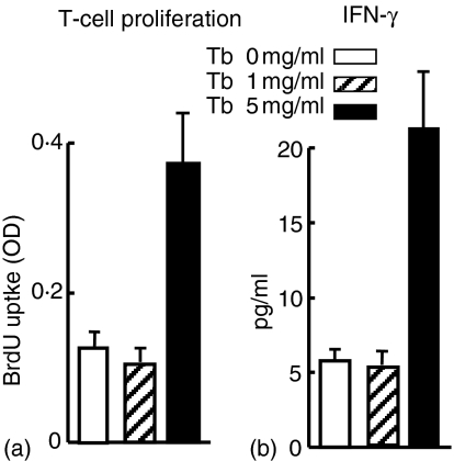
Collagen-induced arthritis (CIA) can be induced even in CIA-resistant H-2(b) background of C57BL/6 mice when these mice are immunized with type II collagen (CII) emulsified in complete Freund's adjuvant (CFA) containing high, but not low, dose of Mycobacterium tuberculosis. Here, we investigated the pathogenesis of CIA in C57BL/6 mice induced by the immunizing protocol. We examined expressions of cytokines, costimulatory molecules and major histocompatibility complex (MHC) class II in draining lymph nodes (DLN) in CIA-induced C57BL/6 mice by quantitative reverse transcription-polymerase chain reaction. We also examined an effect of M. tuberculosis on the expression of these molecules on dendritic cells (DC) in vitro by flow cytometry. We finally examined an effect of M. tuberculosis in CFA used for immunization with CII antigen on priming of CD4+ helper T cells specific to CII in DLN of CIA-induced C57BL/6 mice. The expression of interferon-gamma (IFN-gamma), Interleukin-12p40 (IL-12p40), costimulatory molecules CD40, CD80 and CD86 and MHC class II were up-regulated in DLN of CIA-induced C57BL/6 mice. Expressions of these costimulatory molecules were also up-regulated on DC after stimulation with high, but not low, dose of M. tuberculosis in vitro. Furthermore, priming of CD4+ helper T cells specific to CII antigen in DLN required immunization with CII using CFA containing high, but not low, dose of M. tuberculosis. These results suggested that high dose of M. tuberculosis were required for maturation of DC enough to prime CD4+ helper T cells specific to CII antigen in DLN of H-2b background of C57BL/6 mice.


Figures

Figure 1
Figure 1
Induction of CIA in C57BL/6 mice. C57BL/6 mice were injected i. twice at day −21 and 0 with 100 µg CII emulsified in either IFA (Tb 0 mg/ml) (◊), or CFA containing M. tuberculosis at a concentration of 1 or 5 mg/ml (□ and respectively) (n = 21 in each group). Incidences of CIA and clinical scores were determined, as described in Materials and methods. Data are shown as cumulative incidence (a) and mean clinical score (b) of each group. A hind paw swelling in CIA-induced mouse 10 days after the second immunization and a control non-arthritis paw are shown (c). Representative histological (d) and radiological (e) findings on an affected joint of a CIA-induced C57BL/6 mouse (12 days (d) and 12 weeks (e)) after the second immunization and control joints are also shown (H&E, original magnification ×100).
Figure 2
Figure 2
Expression of cytokines in DLN and affected joints of CIA-induced C57BL/6 mice. mRNA was extracted from DLN (a) and affected joints (b) of CIA-induced C57BL/6 mice 10 days after the second immunization and expression of cytokines indicated were analysed by quantitative RT–PCR analysis. Expressions of these cytokines in corresponding DLN and joints of mice (immunized with CII in IFA) were also shown as controls. Relative expressions of IFN-γ and IL-12 in DLN and joints from CIA-induced C57BL/6 mice significantly increased compared with those of control non-arthritic mice (*; P < 0·05, **; P < 0·01) (n = 10). Data are the means ± SEM of the relative expression of each gene.
Figure 3
Figure 3
Expression of costimulatory molecules and MHC II in DLN of CIA-induced C57BL/6 mice mRNA was extracted from DLN of CIA-induced C57BL/6 mice 10 days after the second immunization and expression of costimulatory molecules indicated were analysed by quantitative RT–PCR analysis. Expressions of these molecules in corresponding DLN of control naive mice were also shown as controls. Relative expressions of CD86, CD40 and MHC II in DLN from CIA-induced C57BL/6 mice significantly increased compared with those of control mice (*P < 0·05) (n = 10). Data are the mean ± SEM of the relative expression of each gene.
Figure 4
Figure 4
Maturation of DC induced by M. tuberculosis in vitro. Splenocytes prepared from naïve C57BL/6 mice were cultured and stimulated or not with 2·5 or 5·0 µg/ml heat-killed M. tuberculosis for 48 hr. Cells were then simultaneously stained with PE-conjugated CD11c and FITC-conjugated monoclonal antibodies against CD80, CD86, CD40 (open histograms) or an isotype control (shaded histograms). Expressions of the costimulatory molecules and MHC II on CD11c+ DC were analysed (a) and mean fluorescence intensities were determined (b) by flow cytometry. Data are representative of four independent experiments.
Figure 5
Figure 5
Maturation of DC induced by M. tuberculosis in vivo. Mice were injected into their foot pads with CII in IFA (Tb; 0 mg/ml) or CFA containing 1 or 5 mg/ml of M. tuberculosis. Five days after the immunization, total RNA was prepared from DLN cells and the expression of each gene was measured by quantitative RT–PCR analysis. Relative expressions of CD86, CD40 and MHC II in DLN from mice immunized with CII with 5 mg/ml of M. tuberculosis were significantly increased compared with those of the other groups (*P < 0·05) (n = 6). Data are the mean ± SEM of the relative expression of each gene.
Figure 6
Figure 6
Collagen specific T cells were recruited to draining lymph nodes in Tb 5 mice. Naïve C57BL/6 mice were immunized with CII antigen emulsified in IFA or CFA containing M. tuberculosis at a final concentration of 1 or 5 mg/ml. Five days after the immunization, CD4+ T cells were purified from DLN and stimulated with denatured chick CII antigen (50 µg/ml) for 48 hr in the presence of mitomycin C-treated splenocytes from naïve C57BL/6 mice as APC. All cultures were pulsed with BrdU for the last 16 hr and CD4+ T-cell proliferation was determined by ELISA. IFN-γ production from CD4+ T cells was also analysed in the culture supernatants by ELISA. Data are the means ± SEM and are representative of three independent experiments.

References

    1. Courtenay JS, Dallman MJ, Dayan AD, Martin A, Mosedale B. Immunisation against heterologous type II collagen induces arthritis in mice. Nature. 1980;283:666–8. - PubMed
    1. Holmdahl R, Andersson ME, Goldschmidt TJ, Jansson L, Karlsson M, Malmstrom V, Mo J. Collagen induced arthritis as an experimental model for rheumatoid arthritis. Immunogenetics, pathogenesis and autoimmunity. Apmis. 1989;97:575–84. - PubMed
    1. Durie FH, Fava RA, Noelle RJ. Collagen-induced arthritis as a model of rheumatoid arthritis. Clin Immunol Immunopathol. 1994;73:11–8. - PubMed
    1. Myers LK, Rosloniec EF, Cremer MA, Kang AH. Collagen-induced arthritis, an animal model of autoimmunity. Life Sci. 1997;61:1861–78. - PubMed
    1. Wooley PH, Luthra HS, Stuart JM, David CS. Type II collagen-induced arthritis in mice. I. Major histocompatibility complex (I region) linkage and antibody correlates. J Exp Med. 1981;154:688–700. - PMC - PubMed

Publication types

MeSH terms