. 2006 Jun 13;34(10):3169-80.
doi: 10.1093/nar/gkl407. Print 2006.

Identifying an interaction site between MutH and the C-terminal domain of MutL by crosslinking, affinity purification, chemical coding and mass spectrometry

Affiliations

Identifying an interaction site between MutH and the C-terminal domain of MutL by crosslinking, affinity purification, chemical coding and mass spectrometry

Robert Ahrends et al. Nucleic Acids Res. .

Abstract

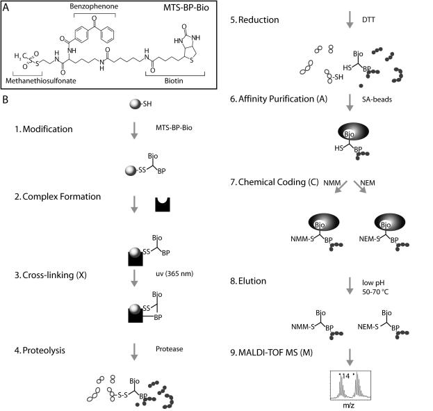
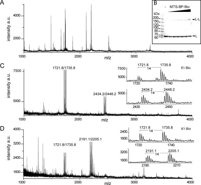
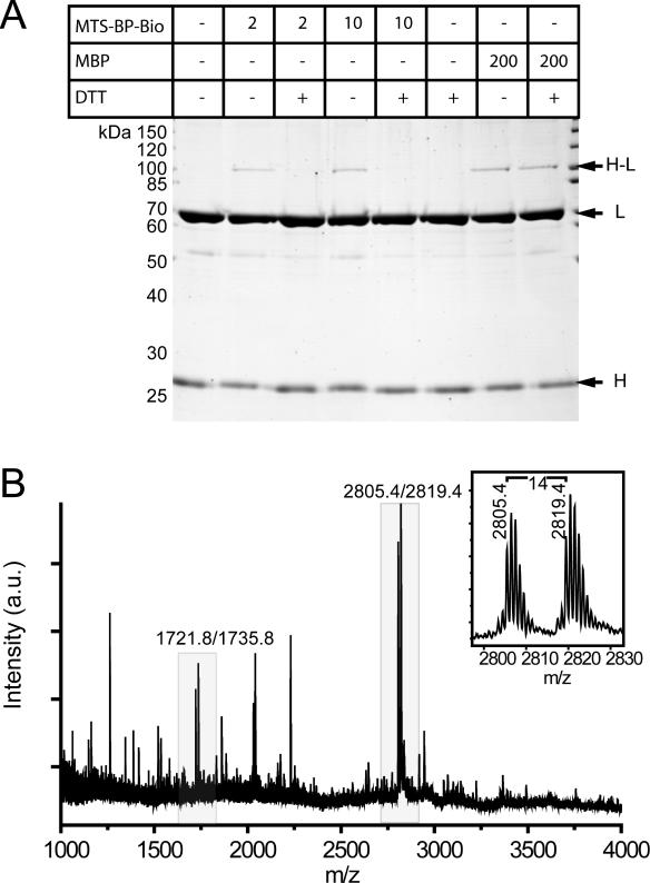
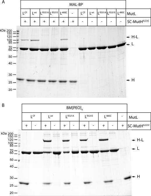
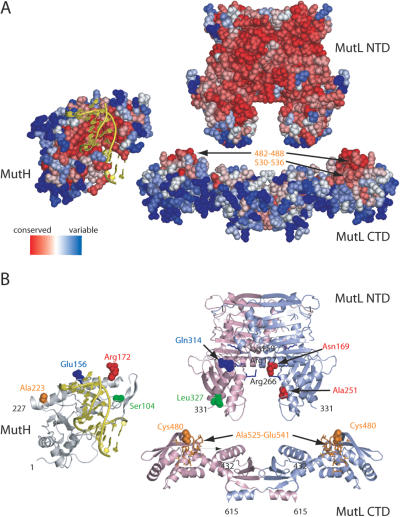
To investigate protein-protein interaction sites in the DNA mismatch repair system we developed a crosslinking/mass spectrometry technique employing a commercially available trifunctional crosslinker with a thiol-specific methanethiosulfonate group, a photoactivatable benzophenone moiety and a biotin affinity tag. The XACM approach combines photocrosslinking (X), in-solution digestion of the crosslinked mixtures, affinity purification via the biotin handle (A), chemical coding of the crosslinked products (C) followed by MALDI-TOF mass spectrometry (M). We illustrate the feasibility of the method using a single-cysteine variant of the homodimeric DNA mismatch repair protein MutL. Moreover, we successfully applied this method to identify the photocrosslink formed between the single-cysteine MutH variant A223C, labeled with the trifunctional crosslinker in the C-terminal helix and its activator protein MutL. The identified crosslinked MutL-peptide maps to a conserved surface patch of the MutL C-terminal dimerization domain. These observations are substantiated by additional mutational and chemical crosslinking studies. Our results shed light on the potential structures of the MutL holoenzyme and the MutH-MutL-DNA complex.


Figures

Figure 1
Figure 1
Schematic representation of XACM (X, Crosslinking; A, affinity purification; C, chemical coding and M, mass spectrometry). (A) Structure of the trifunctional crosslinker MTS-BP-Bio. (B) Schematic representation of the XACM method used to identify protein interaction sites on the peptide level by photocrosslinking/affinity purification/chemical coding/MALDI-TOF MS. (1) A protein is modified at thiol groups with MTS-BP-Bio thereby forming a disulfide bond. (2) The interaction partner is added to allow complex formation (not necessary in case of obligate protein complexes). (3) Photocrosslinking of the benzophenone moiety to the interaction partner is initiated by irradiation at 365 nm. (4) Proteins and crosslinked complexes are then digested in-solution with proteases. (5) The crosslinked products are cleaved at their disulfide linkage thereby transferring the biotin group to the peptide of the interaction partner. (6) The peptides bearing the biotin moiety of the crosslinker are captured on streptavidin coated magnetic beads [14]. (7) The sample is split and the biotinylated peptides are chemically coded at their free thiol group of the crosslinker with N-methylmaleimide (NMM) or N-ethylmaleimide (NEM) to give two products with a mass difference of 14 atomic mass units. (8) The coded peptides are combined and eluted. (9) Coded peptides are identified by MALDI-TOF MS by the characteristic doublets separated by 14 a.m.u.
Figure 2
Figure 2
Analysis of photocrosslinking SC-MutLA282C by SDS–PAGE and MALDI-TOF MS. (A) (Partial positive ion) MALDI-TOF mass spectrum of tryptic peptides (corresponding to 25 pmol input of SC-MutLA282C) before affinity purification. (B) SDS–PAGE analysis (6% gels) of crosslink reactions of homodimeric SC-MutLA282C (5 µM) with increasing molar excess MTS-BP-Bio ranging from 1- to 20-fold. (C) MALDI-TOF spectrum after affinity purification and chemical coding with NMM and NEM. The control peptide K1-Bio (2.5 pmol) was added prior to affinity purification (Table 1). (D) same as (C) but the proteolysis was performed with trypsin and Glu-C. Peaks doublets labeled of the internal biotinylated control peptide K1-Bio coded with NMM and NEM (m/z 1721.8/1735.8), and crosslinked peptides are labeled and shown in the insets (Table 1). All ions are protonated molecules and the m/z values refer to those of monoisotopic mass. Intensities are given in arbitrary units (a.u.).
Figure 3
Figure 3
Analysis of photocrosslinking SC-MutHA223C to its activator protein MutLCF by SDS–PAGE and MALDI-TOF MS. (A) SDS–PAGE analysis of crosslink reactions of the heterocomplex of SC-MutHA223C (5 µM) modified with either 5-fold molar excess of the non-cleavable photocrosslinker MBP in comparison with MTS-BP-Bio. After complex formation with MutLCF and irradiation, (365 nm 10 min) new bands are observed (H-L). (B) Partial positive ion MALDI-TOF mass spectrum of peptides obtained by trypsin/Glu-C digestion of the MTS-BP-Bio crosslinking reaction mixture (corresponding to 25 pmol input of MutL) after affinity purification and chemical coding with NMM and NEM. The control peptide K1-Bio (2.5 pmol) was added prior to affinity purification (Table 1). All ions are protonated molecules and the m/z values refer to those of monoisotopic mass. Intensities are given in arbitrary units (a.u.).
Figure 4
Figure 4
Analysis of crosslinking SC-MutHA223C to its activator protein MutL by SDS–PAGE. (A) Photocrosslink reactions of the heterocomplex formed by SC-MutHA223C modified with MBP and the indicated MutL variants. After complex formation with MutL and UV-irradiation new bands are observed (H–L). Note that both MutLR531A and MutLR531E fail to form a photocrosslink with SC-MutHA223C. (B) Chemical crosslinking of SC-MutHA223C to the indicated MutL variants with the cysteine specific homobifunctional crosslinker BM[PEO]4. All MutL variants but MutLCF are able to form a chemical crosslink with SC-MutHA223C.
Figure 5
Figure 5
Chemical crosslinking SC-MutHA223C to SC-MutL480C abolishes MutS requirement for MutH activation. (A) SDS–PAGE analysis of HPLC-purified complex SC-MutHA223C with SC-MutL480C crosslinked with the cleavable reagent MTS-11-O3-MTS (for details see Materials and Methods) before and after treatment with 5 mM DTT. The amount of uncrosslinked SC-MutHA223C co-purifying with the complex was judged to be <5% of the crosslinked MutH. (B) MutH DNA cleavage promoted by MutL and MutS was assayed as described in Materials and Methods using a 484 bp heteroduplex DNA (25 nM). The complex of SC-MutHA223C/SC-MutL480C (1 µM) crosslinked (−DTT, open symbols) or uncrosslinked (+DTT, closed symbols) was assayed in the absence (triangles) or presence (squares) of 1 µM MutS. Note that only in the crosslinked complex MutH is able to cleave DNA even in the absence of MutS.
Figure 6
Figure 6
Residue sequence conservation and crosslinking results mapped onto the structures of MutH (pdb code 2azo chain B), MutL NTD (pdb code 1b63) and MutL CTD [pdb code 1x9z, (39)]. DNA taken from the structure of the co-crystal from Haemophilus influenzae MutH with specific DNA (pdb code 2aoq) has been superimposed on the structure of E.coli MutH. MutH is shown relative to MutL NTD in an ‘open book’ view based on docking results (11). The NTD and CTD of MutL are aligned arbitrarily to share a common dyad axis. (A) Residue sequence conservation using only protein sequences from bacteria with a gene for both MutH and MutL was obtained using the CONSURF server (56). Conserved residues predicted to be a potential protein interaction site are labeled in yellow (39). (B) Cartoon diagrams of MutH and MutL. The two subunits of the MutL homodimer are colored in light green and blue, respectively. Residues involved in DNA binding (Lys159, Arg177 and Arg266) are shown as sticks colored in blue (49). Positions of cysteine residues in single-cysteine variants of MutH and MutL are indicated as solid spheres. Positions in MutH and MutL that can be crosslink with homobifunctional reagents [Ref. (11) and this study] are shown in the same color. The MutL-peptide crosslinked to cysteine-223 in SC-MutHA223C modified with MTS-BP-Bio and identified using XACM is shown as sticks colored in orange. Note that the conserved site of the CTD should face towards NTD to match all the restraints from the crosslinking experiments (for details see text).

References

    1. Kunkel T.A., Erie D.A. DNA mismatch repair. Annu. Rev. Biochem. 2005;74:681–710. - PubMed
    1. Schofield M.J., Hsieh P. DNA mismatch repair: molecular mechanisms and biological function. Annu. Rev. Microbiol. 2003;57:579–608. - PubMed
    1. Rowley P.T. Inherited susceptibility to colorectal cancer. Annu. Rev. Med. 2005;56:539–554. - PubMed
    1. Modrich P. Mechanisms and biological effects of mismatch repair. Annu. Rev. Genet. 1991;25:229–253. - PubMed
    1. Ban C., Junop M., Yang W. Transformation of MutL by ATP binding and hydrolysis: a switch in DNA mismatch repair. Cell. 1999;97:85–97. - PubMed

Publication types

MeSH terms