. 2006 Oct 1;108(7):2455-62.
doi: 10.1182/blood-2006-04-017251. Epub 2006 Jun 13.

Soluble CD40 ligand accumulates in stored blood components, primes neutrophils through CD40, and is a potential cofactor in the development of transfusion-related acute lung injury

Affiliations

Soluble CD40 ligand accumulates in stored blood components, primes neutrophils through CD40, and is a potential cofactor in the development of transfusion-related acute lung injury

Samina Yasmin Khan et al. Blood. .

Abstract

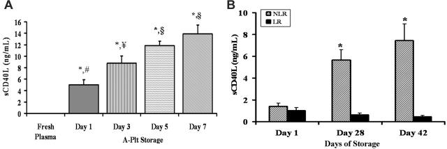
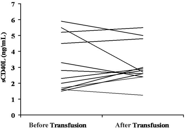
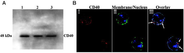
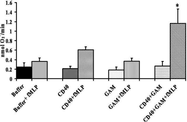
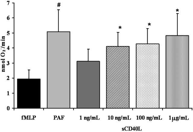
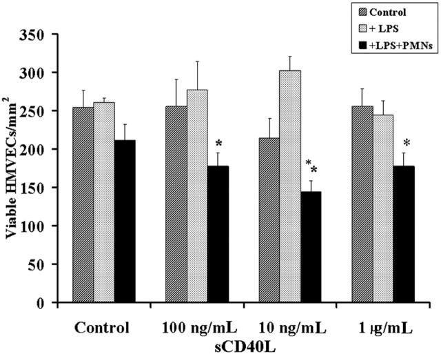
Transfusion-related acute lung injury (TRALI) is a form of posttransfusion acute pulmonary insufficiency that has been linked to the infusion of biologic response modifiers (BRMs), including antileukocyte antibodies and lipids. Soluble CD40 ligand (sCD40L) is a platelet-derived proinflammatory mediator that accumulates during platelet storage. We hypothesized that human polymorpho-nuclear leukocytes (PMNs) express CD40, CD40 ligation rapidly primes PMNs, and sCD40L induces PMN-mediated cytotoxicity of human pulmonary microvascular endothelial cells (HMVECs). Levels of sCD40L were measured in blood components and in platelet concentrates (PCs) implicated in TRALI or control PCs that did not elicit a transfusion reaction. All blood components contained higher levels of sCD40L than fresh plasma, with apheresis PCs evidencing the highest concentration of sCD40L followed by PCs from whole blood, whole blood, and packed red blood cells (PRBCs). PCs implicated in TRALI reactions contained significantly higher sCD40L levels than control PCs. PMNs express functional CD40 on the plasma membrane, and recombinant sCD40L (10 ng/mL-1 mug/mL) rapidly (5 minutes) primed the PMN oxidase. Soluble CD40L promoted PMN-mediated cytotoxicity of HMVECs as the second event in a 2-event in vitro model of TRALI. We concluded that sCD40L, which accumulates during blood component storage, has the capacity to activate adherent PMNs, causing endothelial damage and possibly TRALI in predisposed patients.


Figures

Figure 1.
Figure 1.
Accumulation of sCD40L in stored A-Plts and LR-PRBCs and NLR-PRBCs. (A) A-Plts were serially stored according to AABB standards, plasma samples were obtained and separated from the cells by centrifugation, sCD40L concentration (ng/mL) was measured by commercial ELISA, and fresh, heparinized plasma taken from healthy donors was used as a control for sCD40L concentration. The sCD40L concentration in A-Plts was significantly greater at days 1, 3, 5, and 7 of storage in the A-Plts than in FP (P < .05; n = 5). Moreover, the levels continued to significantly increase on days 3, 5, and 7 compared with day 1. However, the concentrations of sCD40L on day 5 compared with day 7 were not significantly different from one another (P < .05). *Statistical significance from FP (P < .05); ¥,§Significance from day 1 (#) (P < .05). (B) Accumulation of sCD40L in PRBCs in which 50% of the unit (by weight) was leukoreduced (LR) and 50% was unmodified (NLR) before storage. sCD40L concentrations (ng/mL) were measured serially in LR-PRBCs and NLR-PRBCs on days 1, 28, and 42. Day 28 and day 42 NLR (hatched bars) were statistically significant (P < .05; n = 5) compared with day 1 NLR. Days 28 and 42 NLR were also significant from days 1, 28, and 42 LR (solid bars). *Statistical significance (P < .05) compared with day 1 of storage.
Figure 2.
Figure 2.
Soluble CD40L concentration in the plasma of TRALI patients before and after transfusion. Each line reflects the sCD40L in a pretransfusion typing plasma (left) compared with the sCD40L concentration in a plasma sample drawn when TRALI was clinically recognized (right). In 8 of 12 TRALI patients, levels of sCD40L concentrations increased at the time TRALI was diagnosed.
Figure 3.
Figure 3.
Expression of CD40 in human PMNs by Western blot analysis and localization of CD40 by digital microscopy. (A) Experiments were performed on 3 donors. Proteins from PMN whole cell lysates (1.25 × 10⁶) were separated by sodium dodecyl sulfate–polyacrylamide gel electrophoresis (SDS-PAGE) and were immunoblotted with a monoclonal antibody to human CD40. The CD40 glycoprotein band is present at 48 kDa. (B) Fixed PMNs were incubated with a human CD40 monoclonal antibody, and then the images were prepared for digital microscopy as previously described. (i) CD40 immunoreactivity is red because of the secondary donkey antimouse antibody conjugated to Cy3. (ii) The membrane is green because of the association of the wheat germ agglutinin (WGA)–AlexaFluor 488 with salicylic acids, and the nucleus is stained blue with the fluorescent bis-benzimide nuclear dye. (iii) Colocalization of CD40 and the plasma membrane is displayed in pseudocolor. Arrows point to the red area, where a high amount of colocalization is present. Blue represents low colocalization between CD40 and the plasma membrane. These images are representative of 3 different experiments on the PMNs of 3 donors.
Figure 4.
Figure 4.
Cross-linking CD40 primes the PMN oxidase. PMNs were incubated with a mouse monoclonal antibody to human CD40, isotype controls, or buffer for 60 minutes at 4°C, followed by the addition of a buffer control or a goat anti–mouse F(ab′)2 (GAM) to cross-link the CD40 antibody. Activation of the PMN oxidase was accomplished with the addition of 1 μM fMLP, and priming of the fMLP-activated oxidase was calculated as the augmentation of the maximal rate of O2 in response to fMLP in buffer-treated controls. *Significant increase in oxidase activity compared with the buffer-treated controls (P < .05). The only group to significantly prime the fMLP-activated respiratory burst was CD40 + GAM + fMLP, indicating that cross-linking CD40 directly causes priming of the NADPH oxidase. Importantly, incubation of PMNs treated with isotype controls with the F(ab′)2 cross-linkers did not cause priming of the fMLP-activated oxidase (results not shown). This figure is representative of 7 separate experiments using disparate blood donors as the source of the PMNs. Data are presented as mean ± standard error of the mean.
Figure 5.
Figure 5.
Soluble CD40L primes the PMN oxidase. PMN priming assays were completed with incubation of PMNs with buffer, sCD40L (1 ng/mL-1 μg/mL), or PAF (2 μM) (positive control). The ability of sCD40L to prime the fMLP-activated oxidase in human PMNs was measured over a range of concentrations for 5 minutes. Significant priming of the PMN oxidase occurred with the addition of 10 ng/mL, 100 ng/mL, and 1 μg/mL sCD40L compared with buffer-treated controls (#P < .05; *P < .05; n = 7); significant differences exist between groups with different symbols. Data are presented as mean ± standard error of the mean.
Figure 6.
Figure 6.
A 2-event in vitro model of PMN-mediated pulmonary endothelial damage. HMVECs were grown to 80% to 90% confluence on 12-well plates and were incubated with buffer or LPS for 6 hours, followed by the addition of buffer or freshly isolated PMNs that were allowed to settle for 30 minutes at a target-effector ratio of 10:1. After the addition of PMNs or buffer, a range of human recombinant CD40L concentrations or buffer controls was added to the HMVECs. (dark gray bars) Control cells; buffer-treated HMVECs with no PMNs. (light gray bars) HMVECs pretreated with 2 μg/mL LPS for 6 hours at 37°C, followed by treatment with buffer or a range of sCD40L concentrations (as delineated on the x-axis). (black bars) HMVECs incubated with LPS for 6 hours, followed by the addition of PMNs and sCD40L. Incubation of the adherent PMNs with concentrations of sCD40L at 100 ng/mL, 10 ng/mL, and 1 μg/mL resulted in a significant decrease in the number of viable HMVECs. *P < .05 (n = 3) compared with buffer. These data demonstrate that sCD40L may cause concentration-dependent PMN-mediated damage of LPS-activated HMVECs. Importantly, HMVECs stimulated with LPS and exposed to PMNs without sCD40L did not demonstrate PMN-mediated damage but did evidence widespread PMN adherence. Data are presented as mean ± standard error of the mean.

References

    1. Harnett MM. CD40: a growing cytoplasmic tale. Sci STKE. 2004;2004: e25. - PubMed
    1. Inwald DP, McDowall A, Peters MJ, Callard RE, Klein NJ. CD40 is constitutively expressed on platelets and provides a novel mechanism for platelet activation. Circ Res. 2003;92: 1041-1048. - PubMed
    1. Phipps RP, Kaufman J, Blumberg N. Platelet derived CD154 (CD40 ligand) and febrile responses to transfusion. Lancet. 2001;357: 2023-2024. - PubMed
    1. Phipps RP, Koumas L, Leung E, et al. The CD40-CD40 ligand system: a potential therapeutic target in atherosclerosis. Curr Opin Invest Drugs. 2001;2: 773-777. - PubMed
    1. Adawi A, Zhang Y, Baggs R, Finkelstein J, Phipps RP. Disruption of the CD40-CD40 ligand system prevents an oxygen-induced respiratory distress syndrome. Am J Pathol. 1998;152: 651-657. - PMC - PubMed

Publication types