Sclerosing cholecystitis associated with autoimmune pancreatitis
- PMID: 16773691
- PMCID: PMC4087467
- DOI: 10.3748/wjg.v12.i23.3736
Sclerosing cholecystitis associated with autoimmune pancreatitis
Abstract
Aim: To evaluate the histopathological and radiological findings of the gallbladder in patients with autoimmune pancreatitis (AIP).
Methods: The radiological findings of the gallbladder of 19 AIP patients were retrospectively reviewed. Resected gallbladders of 8 AIP patients were examined histologically and were immunostained with anti-IgG4 antibody. Controls consisted of gallbladders resected for symptomatic gallstones (n = 10) and those removed during pancreatoduodenectomy for pancreatic carcinoma (n = 10), as well as extrahepatic bile ducts and pancreases removed by pancreatoduodenectomy for pancreatic carcinoma (n = 10).
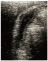
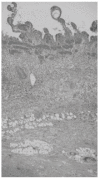
Results: Thickening of the gallbladder wall was detected by ultrasound and/or computed tomography in 10 patients with AIP (3 severe and 7 moderate); in these patients severe stenosis of the extrahepatic bile duct was also noted. Histologically, thickening of the gallbladder was detected in 6 of 8 (75%) patients with AIP; 4 cases had transmural lymphoplasmacytic infiltration with fibrosis, and 2 cases had mucosal-based lymphoplasmacytic infiltration. Considerable transmural thickening of the extrahepatic bile duct wall with dense fibrosis and diffuse lymphoplasmacytic infiltration was detected in 7 patients. Immunohistochemically, severe or moderate infiltration of IgG4-positive plasma cells was detected in the gallbladder, bile duct, and pancreas of all 8 patients, but was not detected in controls.
Conclusion: Gallbladder wall thickening with fibrosis and abundant infiltration of IgG4-positive plasma cells is frequently detected in patients with AIP. We propose the use of a new term, sclerosing cholecystitis, for these cases that are induced by the same mechanism as sclerosing pancreatitis or sclerosing cholangitis in AIP.
Figures


References
-
- Kim KP, Kim MH, Song MH, Lee SS, Seo DW, Lee SK. Autoimmune chronic pancreatitis. Am J Gastroenterol. 2004;99:1605–1616. - PubMed
-
- Kamisawa T, Egawa N, Nakajima H, Tsuruta K, Okamoto A. Morphological changes after steroid therapy in autoimmune pancreatitis. Scand J Gastroenterol. 2004;39:1154–1158. - PubMed
-
- Hirano K, Shiratori Y, Komatsu Y, Yamamoto N, Sasahira N, Toda N, Isayama H, Tada M, Tsujino T, Nakata R, et al. Involvement of the biliary system in autoimmune pancreatitis: a follow-up study. Clin Gastroenterol Hepatol. 2003;1:453–464. - PubMed
-
- Kamisawa T, Funata N, Hayashi Y, Eishi Y, Koike M, Tsuruta K, Okamoto A, Egawa N, Nakajima H. A new clinicopathological entity of IgG4-related autoimmune disease. J Gastroenterol. 2003;38:982–984. - PubMed
Publication types
MeSH terms
Substances
LinkOut - more resources
Full Text Sources
Medical
Miscellaneous

