

Cholangiocytes and blood supply
- PMID: 16773711
- PMCID: PMC4087570
- DOI: 10.3748/wjg.v12.i22.3546
Cholangiocytes and blood supply
Abstract
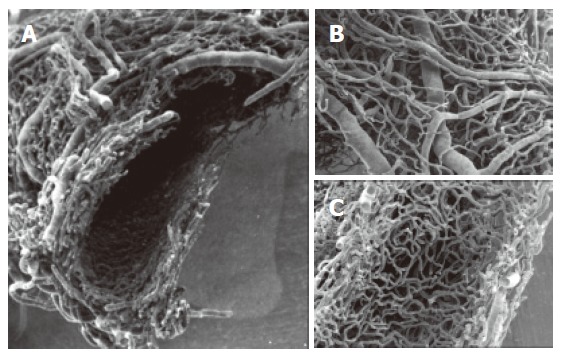
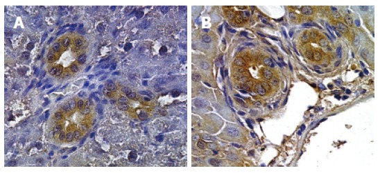
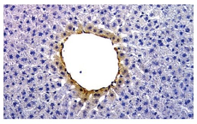
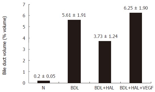
The microvascular supply of the biliary tree, the peribiliary plexus (PBP), stems from the hepatic artery branches and flows into the hepatic sinusoids. A detailed three-dimensional study of the PBP has been performed by using the Scanning Electron Microscopy vascular corrosion casts (SEMvcc) technique. Considering that the PBP plays a fundamental role in supporting the secretory and absorptive functions of the biliary epithelium, their organization in either normalcy and pathology is explored. The normal liver shows the PBP arranged around extra- and intrahepatic biliary tree. In the small portal tract PBP was characterized by a single layer of capillaries which progressively continued with the extrahepatic PBP where it showed a more complex vascular network. After common duct ligation (BDL), progressive modifications of bile duct and PBP proliferation are observed. The PBP presents a three-dimensional network arranged around many bile ducts and appears as bundles of vessels, composed by capillaries of homogeneous diameter with a typical round mesh structure. The PBP network is easily distinguishable from the sinusoidal network which appears normal. Considering the enormous extension of the PBP during BDL, the possible role played by the Vascular Endothelial Growth Factor (VEGF) is evaluated. VEGF-A, VEGF-C and their related receptors appeared highly immunopositive in proliferating cholangiocytes of BDL rats. The administration of anti-VEGF-A or anti-VEGF-C antibodies to BDL rats as well as hepatic artery ligation induced a reduced bile duct mass. The administration of rVEGF-A to BDL hepatic artery ligated rats prevented the decrease of cholangiocyte proliferation and VEGF-A expression as compared to BDL control rats. These data suggest the role of arterial blood supply of the biliary tree in conditions of cholangiocyte proliferation, such as it occurs during chronic cholestasis. On the other hand, the role played by VEGF as a tool of cross-talk between cholangiocytes and PBP endothelial cells suggests that manipulation of VEGF release and function could represent a therapeutic strategy for human pathological conditions characterized by damage of hepatic artery or the biliary tree.
Figures


References
-
- Tietz PS, LaRusso NF. Cholangiocyte biology. Curr Opin Gastroenterol. 2005;21:337–343. - PubMed
-
- Alvaro D. Biliary epithelium: a new chapter in cell biology. Ital J Gastroenterol Hepatol. 1999;31:78–83. - PubMed
-
- Demetris AJ. Participation of cytokines and growth factors in biliary cell proliferation and mito-inhibition during ductular reaction. In: Alpini G, Alvaro D, LeSage G, Marzioni M, LaRusso NF, et al., editors. Pathophysiology of the Bile Duct System. Georgetown, Texas, USA: Landes Biosciences; 2004. pp. 167–182.
-
- Strazzabosco M, Fabris L, Spirli C. Pathophysiology of cholangiopathies. J Clin Gastroenterol. 2005;39:S90–S102. - PubMed
-
- Ohtani O, Kikuta A, Ohtsuka A, Taguchi T, Murakami T. Microvasculature as studied by the microvascular corrosion casting/scanning electron microscope method. I. Endocrine and digestive system. Arch Histol Jpn. 1983;46:1–42. - PubMed
Publication types
MeSH terms
Substances
Grants and funding
LinkOut - more resources
Full Text Sources
Medical
Research Materials
Miscellaneous
