. 2006 Jul;4(7):e229.
doi: 10.1371/journal.pbio.0040229.

AgDscam, a hypervariable immunoglobulin domain-containing receptor of the Anopheles gambiae innate immune system

Affiliations

AgDscam, a hypervariable immunoglobulin domain-containing receptor of the Anopheles gambiae innate immune system

Yuemei Dong et al. PLoS Biol. 2006 Jul.

Abstract

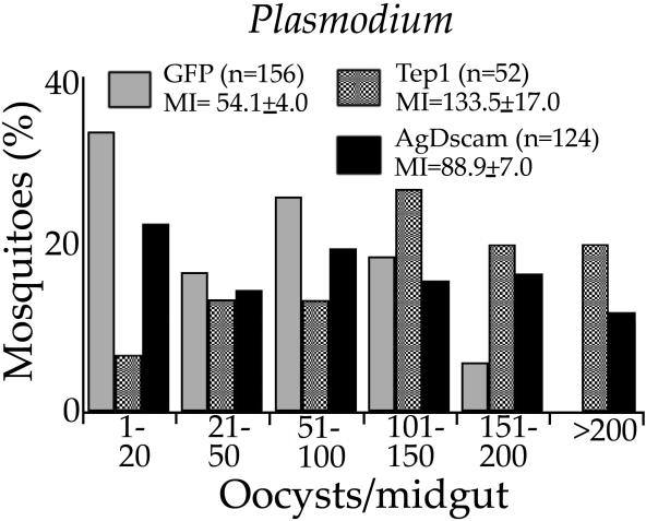
Activation of the insect innate immune system is dependent on a limited number of pattern recognition receptors (PRRs) capable of interacting with pathogen-associated molecular pattern. Here we report a novel role of an alternatively spliced hypervariable immunoglobulin domain-encoding gene, Dscam, in generating a broad range of PRRs implicated in immune defense in the malaria vector Anopheles gambiae. The mosquito Down syndrome cell adhesion molecule gene, AgDscam, has a complex genome organization with 101 exons that can produce over 31,000 potential alternative splice forms with different combinations of adhesive domains and interaction specificities. AgDscam responds to infection by producing pathogen challenge-specific splice form repertoires. Transient silencing of AgDscam compromises the mosquito's resistance to infections with bacteria and the malaria parasite Plasmodium. AgDscam is mediating phagocytosis of bacteria with which it can associate and defend against in a splice form-specific manner. AgDscam is a hypervariable PRR of the A. gambiae innate immune system.


Figures

Figure 1
Figure 1. AgDscam Gene Organization and Infection-Responsive Splicing
(A) The variable Ig domain exon cassettes 4, 6, and 10 are displayed as Ig4(14) (red), Ig6(30) (blue), and Ig10(38) (green), each containing 14, 30, and 38 exons, respectively. The transmembrane domain exons (tm) 11–13 are displayed in pink, and non-spliced exons are displayed in black. (B) Percentage identity of AgDscam exons to the D. melanogaster Dscam exons at the amino acid level is displayed. Phylogenetic relations between AgDscam Ig exons are presented in Figure S1. (C) Differences in AgDscam exon 4 transcript representations between bacterially- (E. coli, P. veronii, S. aureus), fungus- (B. bassiana), LPS-, and PG-challenged cell line Sua5B and a non-challenged cell line at 12 h after challenge, and Plasmodium- (P. berghei and P. falciparum) infected midguts (Mosq) and non-infected blood-fed midguts at 24 h after ingestion. The numbers (“4.1”–“4.14”) indicate the individual exons of exon cassette 4. Expression was determined with RTqPCR analyses in three replica assays. The fold difference in exon representation (ratio) between challenged and naive samples from three replicates were determined in normalized cDNAs, and expression ratios are displayed in a color scheme where red indicates a higher representation and green indicates a lower representation of exons in challenged samples compared to naïve samples. Black was indicative of a lack of infection-responsive regulation. Normalization was done with expression analysis of a non-spliced AgDscam exon and a ribosomal S7 gene. The significance of variable exon regulation at a 95% confidence level, the RTqPCR efficiencies for each pair of exon primers, expression data values, and standard errors are presented in Table S1.
Figure 2
Figure 2. AgDscam Is Implicated in Anti-Bacterial Defense
(A) RNAi-mediated depletion of AgDscam with a dsRNA fragment complementary to a non-spliced AgDscam exon resulted in decreased mosquito survival after challenge with S. aureus and E. coli compared to the challenged control GFP dsRNA-treated (GFP) mosquitoes. AgDscam dsRNA-treated mosquitoes with a significant decrease in survival compared to GFP dsRNA-treated (GFP) mosquitoes, according to a 2-way ANOVA ( p < 0.01), are indicated with asterisks. A Kaplan Meier survival analysis of GFP dsRNA- and AgDscam dsRNA-treated mosquitoes is presented in Figure S3. PBS-injected GFP dsRNA- and AgDscam dsRNA-treated mosquitoes (“GFP + PBS” and “AgDscam + PBS”) did not exhibit significant mortality. The survival rates of AgDscam dsRNA-treated mosquitoes and Gambicin dsRNA-treated mosquitoes after injection with Gram-positive bacteria (S. aureus) were similar. (B) RNAi-mediated depletion of AgDscam in absence of experimental bacterial challenge resulted in a proliferation of opportunistic bacteria in the mosquito haemolymph at 4 d after dsRNA injection, compared to non-treated mosquitoes (naïve) and control GFP dsRNA-treated mosquitoes (GFP). The proliferating bacterial species, capable of growing on LB agar, were determined as species closely related to Bacterium HPC1068, Asaia bogorensis (A.b. ), and P. veronii (P.v.). The total number of bacteria isolated from the haemolymph is indicated with a separate bar (total). (C) Western analysis of AgDscam RNAi-treated 4-d-old female mosquitoes (KD) (mosquito) showed decreased AgDscam protein (DS) by approximately 50% compared to dsGFP-treated control (GFP). Total protein was extracted 4 d after dsRNA injection and normalized for equal actin (Act) content.
Figure 3
Figure 3. AgDscam Is Implicated in Anti- Plasmodium Defense
RNAi-mediated depletion of AgDscam from adult female mosquitoes resulted in increased permissiveness to P. berghei infection, as indicated by a 59%–99% increase in oocysts numbers in four independent assays. The figure presents the frequency distributions of oocysts pooled from four independent assays where MI indicates mean intensity of infection (oocysts number) plus/minus standard error, and n indicates the total number of mosquitoes in each experiment. Infection levels in AgDscam-silenced mosquitoes were comparable to the positive control Tep1 dsRNA-treated mosquitoes [ 44].
Figure 4
Figure 4. AgDscam Is Implicated in Phagocytosis of Bacteria
(A) AgDscam distribution in DAPI-stained Sua5B cells and co-localization with FITC-labeled bacteria [ 30]. In this figure, (a) AgDscam (red) was evenly distributed on the cell surface of non-challenged cells. (b, c1–c3) FITC-labeled E. coli (green, c1) that were co-incubated with the cell line co-localized with AgDscam (red, c2; white, b). Co-localization is indicated in white (b and c3) where cell nuclei were stained with DAPI (blue). (d1–d3) Incubation of FITC-labeled S. cerevisiae (d1) with the same cell line (d2) did not result in phagocytosis and co-localization with AgDscam (d3). S. cerevisiae did not adhere to the cells. Controls: FITC-labeled E. coli (e1) did not interact with the AgDScam antibody or the secondary antibody only (e2). Scale bars: 10 μm. (B) Western analysis of AgDscam (Ds) RNAi-treated Sua5B cell culture (KD) showed decreased AgDscam protein by approximately 82% compared to dsGFP-treated control cells (GFP). Total protein was extracted after 6 d of dsRNA treatments and samples were normalized for equal actin (Act) content. (C) Phagocytic assay of AgDscam-depleted (AgDscam) and control GFP dsRNA-treated Sua5B cells. AgDscam depletion resulted in approximately 50% reduction of phagocytic index for both E. coli and S. aureus, which were significantly different from GFP controls. Asterisks indicate significant decrease at p < 0.01 ( E. coli: t-test, p = 0.006, df = 36; S. aureus: t-test, p = 0.008, df = 40). The phagocytic index was calculated as the ratio of the number of immune competent cells containing fluorescent bacteria against the total number of cells in each field. For each assay at least 20 fields were included and data here represented the mean value from three independent assays with standard error bars included.
Figure 5
Figure 5. Pathogen-Induced AgDscam Splice Form Repertoires Have Increased Affinity to, and Defense Activity against, the Eliciting Pathogen
(A) E. coli and P. veronii had increased affinity to both membrane-bound (M) and secreted (S) AgDscam splice form repertoires that were produced by Sua5B cells previously challenged with E. coli (E.c.) or P. veronii (P.v.), respectively, compared to cells challenged with S. aureus (S.a), or control cells treated with PBS. Similarly, P. veronii showed increased affinity to AgDscam splice forms that were produced by cells previously challenged with P. veronii, compared to cells challenged with E. coli. AgDscam was eluted from the bacteria surface with high salt concentrations (E4 = 400 mM, E6 = 500 mM +100 mM NH 4Ac) after co-incubation with a cell line Sua5B membrane or secreted protein extract followed by washes with PBS. The lower degree of pathogen-specificity for the membrane-bound AgDscam form at the E4 elution was most likely due to excessive amount of bound protein to the bacteria surface. The last elution of the highest stringency was expected to release the AgDscam splice forms with the highest affinity and specificity to the bacteria. (B) Silencing of pathogen-induced specific isoform repertoires affect binding of AgDscam to the inducing pathogen. Silencing of the E. coli-induced exon 4.8-containing splice forms (4.8) resulted in decreased binding of AgDscam to E. coli and P. veronii compared to GFP and exon 4.1 dsRNA-treated cells, while silencing of the S. aureus-induced exon 4.1 (4.1) did not affect AgDscam binding to the two Gram-negative bacteria. This assay was done with both non-challenged cells (PBS) and cells that had been challenged 2 d after dsRNA treatments (GFP, Ds, 4.1, 4.8). Silencing of the total AgDscam (Ds) abolished AgDscam binding to both bacteria species. (C) Pathogen-induced splice form repertoires display increased defense activity to the eliciting pathogen. Upon selective silencing of the E. coli-induced splice form repertoire containing exon 4.8, mosquito's survival rate after G (E. coli) challenge was significantly lower than that for G + (S. aureus) challenge (2-way ANOVA, p < 0.05). Conversely, selective silencing of the S. aureus-induced splice form repertoire containing exon 4.1 rendered the mosquitoes more sensitive to challenge with S. aureus (2-way ANOVA, p < 0.05). GFP dsRNA-treated mosquitoes challenged with E. coli (GFP G−) or S. aureus (GFP G+), total AgDscam dsRNA-treated mosquitoes challenged with E. coli (Ds G ) or S. aureus (Ds G +), exon 4.1 dsRNA-treated mosquitoes challenged with E. coli (4.1 G ) or S. aureus (4.1 G +), exon 4.8 dsRNA-treated mosquitoes challenged with E. coli (4.8 G ) or S. aureus (4.8 G +). (D) RTqPCR validation of RNAi gene-silencing efficiency and specificity. dsRNAs or siRNAs were used to target and specifically silence all AgDscam transcripts (AgDscam) and exon 4.1-, and exon 4.8-containing transcripts (displayed on the x-axis). The efficacy of silencing was assayed by RTqPCR with the primers specific for exon 4.1 (4.1), exon 4.8 (4.8), or the constant Dscam (Ds) for determination of the respective transcript abundance in the different gene-silenced samples and in a control GFP dsRNA-treated mosquito sample. The fold change of the expression is presented as the percentage (%) change compared to the dsGFP-treated control samples. cDNA template amounts from the different samples were normalized through amplification of an A. gambiae ribosomal S7 gene fragment as previously described [ 30]. Standard error bars from the three replica assays are presented.

Comment in

References

    1. Govind S, Nehm RH. Innate immunity in fruit flies: A textbook example of genomic recycling. PLoS Biol. 2004;2:e276. doi: 10.1371/journal.pbio.0020276. - DOI - PMC - PubMed
    1. Hultmark D. Drosophila immunity: Paths and patterns . Curr Opin Immunol. 2003;15:12–19. - PubMed
    1. Meister M. Blood cells of Drosophila Cell lineages and role in host defense . Curr Opin Immunol. 2004;16:10–15. - PubMed
    1. Dimopoulos G. Insect immunity and its implication in mosquito-malaria interactions. Cell Microbiol. 2003;5:3–14. - PubMed
    1. Hoffmann JA. The immune response of Drosophila . Nature. 2003;426:33–38. - PubMed

Publication types

MeSH terms

Associated data