. 2006 Sep;17(9):3717-28.
doi: 10.1091/mbc.e06-03-0244. Epub 2006 Jun 14.

Gab1 is required for cell cycle transition, cell proliferation, and transformation induced by an oncogenic met receptor

Affiliations

Gab1 is required for cell cycle transition, cell proliferation, and transformation induced by an oncogenic met receptor

Kathleen Mood et al. Mol Biol Cell. 2006 Sep.

Abstract

We have shown previously that either Grb2- or Shc-mediated signaling from the oncogenic Met receptor Tpr-Met is sufficient to trigger cell cycle progression in Xenopus oocytes. However, direct binding of these adaptors to Tpr-Met is dispensable, implying that another Met binding partner mediates these responses. In this study, we show that overexpression of Grb2-associated binder 1 (Gab1) promotes cell cycle progression when Tpr-Met is expressed at suboptimal levels. This response requires that Gab1 possess an intact Met-binding motif, the pleckstrin homology domain, and the binding sites for phosphatidylinositol 3-kinase and tyrosine phosphatase SHP-2, but not the Grb2 and CrkII/phospholipase Cgamma binding sites. Importantly, we establish that Gab1-mediated signals are critical for cell cycle transition promoted by the oncogenic Met and fibroblast growth factor receptors, but not by progesterone, the natural inducer of cell cycle transition in Xenopus oocytes. Moreover, Gab1 is essential for Tpr-Met-mediated morphological transformation and proliferation of fibroblasts. This study provides the first evidence that Gab1 is a key binding partner of the Met receptor for induction of cell cycle progression, proliferation, and oncogenic morphological transformation. This study identifies Gab1 and its associated signaling partners as potential therapeutic targets to impair proliferation or transformation of cancer cells in human malignancies harboring a deregulated Met receptor.


Figures

Figure 1.
Figure 1.
Tpr-Met mutants that induce GVBD have the capacity to recruit Gab1. Diagram displays the mechanism by which each mutant of the Tpr-Met oncoprotein is predicted to recruit the Gab1 protein and whether these Tpr-Met oncoproteins induce GVBD (Mood et al., 2006): Tpr-Met, wt Tpr-Met oncoprotein, which interacts with Gab1 via direct and indirect Grb2-dependent mechanisms (Lock et al., 2003); Y482F, mutant of Tpr-Met where Tyr-482 is replaced with Phe (Fixman et al., 1996) and cannot bind directly to Gab1 but retains a Grb2-dependent means to associate with Gab1 (Lock et al., 2003); Y482/489F, mutant of Tpr-Met where Tyr-482 and Tyr-489 are replaced with Phe (Fixman et al., 1996), preventing direct and indirect interaction with Gab1; Shc1, Shc-specific docking Tpr-Met variant that interacts with Gab1 via a Grb2-dependent mechanism (Saucier et al., 2002); Grb2, Grb2-specific docking Tpr-Met variant that interacts indirectly with Gab1 (Saucier et al., 2002); N491H, mutant of Tpr-Met where Gln-491 is replaced with His, preventing direct binding of Grb2 (Fixman et al., 1996), but it can interact directly with Gab1 via phosphotyrosine Y482 (Lock et al., 2003); PTB, PTB domain of Shc, which when expressed in cells, blocks the association of Tpr-Met with the adaptor protein Shc (Mood et al., 2006); and Gab1-Grb2–associated binder 1 and MBM-Met receptor binding motif of Gab1 (Lock et al., 2003).
Figure 2.
Figure 2.
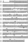
Xenopus, mouse, and human Gab1 proteins are structurally conserved. The putative full-length Gab1 cDNA was cloned by PCR from a Xenopus oocyte cDNA library. The alignment of XGab1 amino acid sequences with those of the human and mouse Gab1 proteins is shown (Holgado-Madruga et al., 1996; Weidner et al., 1996). Conserved residues are highlighted in gray; the PH domain and MBD are denoted by a full or dashed underline, respectively. Lines above the sequences indicate the conserved PI3K, Crk, PLCγ, RasGAP, or SHP-2 binding sites. Potential ERK phosphorylation sites are denoted by lines below the sequences. The Grb2 carboxy-terminal SH3 domain binding sites and the MBM are marked by underbracket and a dotted underline, respectively.
Figure 3.
Figure 3.
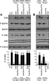
Gab1 is sufficient to mediate cell cycle progression and activation of MAPK and JNK by low levels of the Tpr-Met oncoprotein. (A) Oocytes were injected with suboptimal levels of Tpr-Met, Y482F, or N491H RNA (80 pg/oocyte) in the presence or absence of Gab1 (400 pg/oocyte) and ShcPTB (400 pg/oocyte). Oocytes left uninjected or treated with 5 μM progesterone were used, respectively, as negative and positive controls for induction of GVBD. (B) Oocytes were injected with low (80 pg/oocyte) or high (400 pg/oocyte) concentrations of the Tpr-Met Shc-specific docking variant RNA in the presence or absence of Gab1 (400 pg/oocyte), or ShcPTB (400 pg/oocyte) RNA. Histograms show the percentage of oocytes displaying GVBD, and the number of oocytes with GVBD over the number of oocytes injected is indicated above bars. Lysates prepared from oocytes described in A and B were analyzed by immunoblot conducted with specific antibodies raised against Gab1, phospho-MAPK, MAPK, phospho-JNK, JNK1/2, or phospho-tyrosine15-Cdc2. These data are representative of three independent experiments. Un, uninjected; Pro, treated with 5 μM progesterone; Gab, mouse Gab1; Shc1, the Tpr-Met Shc-specific docking variant (Saucier et al., 2002); and ShcPTB, Myc-tagged Shc PTB domain (Mood et al., 2006).
Figure 4.
Figure 4.
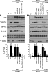
The Grb2 binding site of Gab1, but not the MBM, is dispensable for the induction of cell cycle progression and activation of MAPK and JNK by the Tpr-Met oncoprotein. (A) Suboptimal levels of Tpr-Met RNA (80 pg/oocyte) were injected in oocytes in the presence or absence of RNAs encoding wt or mutant forms of Gab1 (400 pg/oocyte). Oocytes left uninjected or treated with 5 μM progesterone were used, respectively, as negative and positive controls for GVBD. (B) Oocytes were injected with low levels of the N491H Tpr-Met mutant RNA (80 pg/oocyte) in the presence or absence of the wt or mutant forms of Gab1 RNAs (400 pg/oocyte), or they were singly injected with optimal concentrations of the Tpr-Met Shc-specific docking variant RNA (400 pg/oocyte), in the presence or absence of the dominant-negative ShcPTB RNA (400 pg/oocyte). These data are representative of three independent experiments, and the number of oocytes displaying GVBD over the number of oocytes injected is shown as a percentage in histograms. Lysates prepared from oocytes described above were analyzed by immunoblot conducted with specific antibodies raised against Gab1, phospho-MAPK, MAPK, phospho-JNK, or JNK1/2 or phospho-tyrosine15-Cdc2. GabΔGrb2, mutant of Gab1 with deletion of proline-rich domains (Pro4, aa 337-346/Pro5, aa 517-522) required for Grb2 interaction (Lock et al., 2000); GabΔMBM, mutant of Gab1 where Arg-491 is substituted with Ala, disrupting the MBM essential for direct interaction with the Met receptor (Lock et al., 2003); and GabΔMBMΔGrb2, mutant of Gab1 lacking both the Grb2 binding sites and MBM (Lock et al., 2003).
Figure 5.
Figure 5.
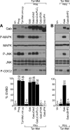
Cell cycle progression mediated by Gab1 requires an intact PH domain, PI3K and SHP-2 binding sites, but not the Grb2 or Crk/PLCγ binding sites. (A and B) Suboptimal levels of Tpr-Met RNA (80 pg/oocyte) were injected in oocytes in the presence or absence of wt or mutant forms of Gab1 RNAs (400 pg/oocyte). Oocytes left uninjected or treated with 5 μM progesterone were used as negative and positive controls for GVBD, respectively. The histograms represent the number of oocytes displaying GVBD over the number of oocytes injected. The data are representative of three independent experiments. Oocyte lysates were analyzed by immunoblot conducted with specific antibodies raised against Gab1, phospho-MAPK, MAPK, phospho-JNK, JNK1/2 or phospho-tyrosine15-Cdc2. GabΔCrkΔPLCγ, mutant of Gab1 where Tyr-242, -265, -307, -317, -373, and -404 are replaced with Phe, preventing the binding of Crk and PLCγ (Lamorte et al., 2000); GabΔPH, mutant of Gab1 deleted of amino acids 1-115 encoding the PH domain (Maroun et al., 1999a); GabΔPI3K, mutant of Gab1 where Tyr-449, -474, and -591 are replaced with Phe, preventing the recruitment of PI3K (Holgado-Madruga et al., 1997); and GabΔSHP2, mutant of Gab1 where Tyr-659 is substituted with Phe, disrupting the binding of the tyrosine SHP-2 (Maroun et al., 2000).
Figure 6.
Figure 6.
The Gab1 MBD interacts with Tpr-Met and impairs Gab1 phosphorylation. (A and B) Independent transient transfection of HEK 293 cells with Tpr-Met or/and HA-tagged wt MBD of Gab1, or a mutant of lacking the Grb2- and Met-binding motifs. (A) Top, Tpr-Met proteins were immunoprecipitated and subsequently blotted with phosphotyrosine antibody to detect phosphorylated Tpr-Met and then stripped and reblotted for detection of Tpr-Met using antibody 144, or blotted with HA for detection of the MBD proteins. Bottom, Western analyses of total cell lysates (TCL) show the levels of HA-tagged MBD or tubulin expression. (B) Top, Tpr-Met proteins were immunoprecipitated and blotted with Tpr-Met–specific antibody. Bottom, Western analyses of TCL show the expression level of HA-tagged MBD, phospho-Gab1, Gab1 proteins, or tubulin.
Figure 7.
Figure 7.
The Gab1 MBD represses cell cycle progression mediated by the oncogenic Met receptor. Oocytes were injected with optimal levels of Tpr-Met RNA (500 pg/oocyte) in the presence or absence of RNAs encoding wt or mutants forms of the Gab1 MBD (14 ng/oocyte). Histograms show the percentage of oocytes displaying GVBD, and the number of oocytes with visible GVBD over the number of oocytes injected is indicated above histogram bars. The data are representative of three independent experiments. Lysates prepared from oocytes were analyzed by immunoblot conducted with antibodies raised against the Met receptor, HA to detect HA-tagged MBD, phospho-MAPK, MAPK, phospho-JNK, or JNK1/2. MBD, wt, MBD of Gab1; MBDΔPI3K, mutant of the Gab1 MBD lacking the PI3K binding sites; MBDΔGrb2, mutant of the Gab1 MBD lacking Grb2 interaction site; MBDΔMBM, mutant of the Gab1 MBD lacking the MBM; and MBDΔMBMΔGrb2, mutant of the Gab1 MBD lacking the Grb2 and Met receptor binding sites.
Figure 8.
Figure 8.
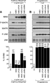
The MBD of Gab1 represses FGFR1-mediated cell cycle, but not GVBD induced by progesterone. (A) Oocytes injected with FGFR1 and FRS2 mRNAs (4 ng each/oocyte) in the presence or absence of wt or mutant of MBD of Gab1 mRNAs (14 ng/oocyte) were stimulated with 200 ng/ml human recombinant basic FGF. (B) Oocytes left uninjected or injected with wt or mutant MBD mRNAs (14 ng/oocyte) were stimulated with 5 μM progesterone. Histograms represent the number of oocytes with visible GVBD over the number of oocytes injected and shown as percentage of GVBD. The data are representative of three independent experiments. Lysates prepared from oocytes were analyzed by immunoblot conducted with specific antibodies raised against HA, to detect HA-tagged MBD, phospho-MAPK, MAPK, phospho-JNK, or JNK1/2.
Figure 9.
Figure 9.
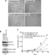
The MBD of Gab1 is sufficient to reverse Tpr-Met–mediated cell transformation and proliferation. (A) Representative morphology of parental Fr3T3 or Tpr-Met–expressing Fr3T3 fibroblast cells infected with retroviruses encoding the indicated Gab1 MBD constructs. Photographs were taken at a 10× magnification after retroviral infection of Tpr-Met–expressing cells with the Gab1 MBD, the MBDΔMBMΔGrb2, or empty vector, 2 wk after selection with 2 μg/ml puromycin. (B) Expression levels of the MBD or Tpr-Met protein in Fr3T3 stable cell lines. Top, Tpr-Met proteins were immunoprecipitated from lysates of Fr3T3 stable cell lines expressing the HA-tagged MBD and Tpr-Met. The expression of Tpr-Met protein and its level of phosphorylation on tyrosine residues are shown. Bottom, level of MBD and tubulin proteins expressed in each cell line by Western analyses conducted with HA or tubulin-specific antibodies. (C) Cell proliferation assays were performed with the indicated stable cell lines. Data on the graph represents the number of cells ± the mean SD (n = 3) over time (hour), and the doubling time was estimated from the curves. Similar results were obtained with other independent cell lines.

Similar articles

Cited by

References

    1. Bardelli A., Basile M. L., Audero E., Giordano S., Wennstrom S., Menard S., Comoglio P. M., Ponzetto C. Concomitant activation of pathways downstream of Grb2 and PI 3-kinase is required for MET-mediated metastasis. Oncogene. 1999;18:1139–1146. - PubMed
    1. Bardelli A., Longati P., Gramaglia D., Stella M. C., Comoglio P. M. Gab1 coupling to the HGF/Met receptor multifunctional docking site requires binding of Grb2 and correlates with the transforming potential. Oncogene. 1997;15:3103–3111. - PubMed
    1. Birchmeier C., Birchmeier W., Gherardi E., Vande Woude G. F. Met, metastasis, motility and more. Nat. Rev. Mol. Cell Biol. 2003;4:915–925. - PubMed
    1. Bladt F., Riethmacher D., Isenmann S., Aguzzi A., Birchmeier C. Essential role for the c-met receptor in the migration of myogenic precursor cells into the limb bud. Nature. 1995;376:768–771. - PubMed
    1. Browaeys-Poly E., Cailliau K., Vilain J. P. Signal transduction pathways triggered by fibroblast growth factor receptor 1 expressed in Xenopus laevis oocytes after fibroblast growth factor 1 addition. Role of Grb2, phosphatidylinositol 3-kinase, Src tyrosine kinase, and phospholipase Cgamma. Eur. J. Biochem. 2000;267:6256–6263. - PubMed

Publication types

MeSH terms

Substances