. 2006 Sep;17(9):3729-44.
doi: 10.1091/mbc.e05-11-1083. Epub 2006 Jun 14.

Phosphoinositide 3-Kinase C2beta regulates cytoskeletal organization and cell migration via Rac-dependent mechanisms

Affiliations

Phosphoinositide 3-Kinase C2beta regulates cytoskeletal organization and cell migration via Rac-dependent mechanisms

Roy M Katso et al. Mol Biol Cell. 2006 Sep.

Abstract

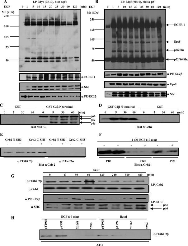
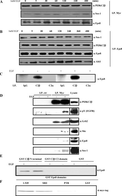
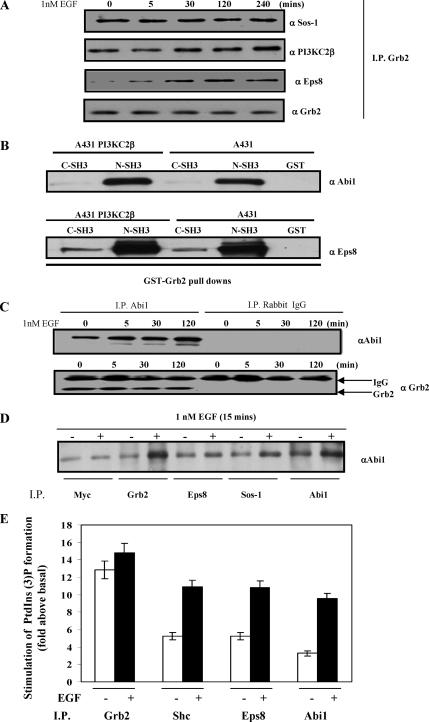
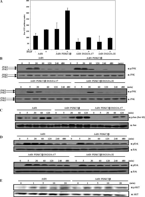
Receptor-linked class I phosphoinositide 3-kinases (PI3Ks) induce assembly of signal transduction complexes through protein-protein and protein-lipid interactions that mediate cell proliferation, survival, and migration. Although class II PI3Ks have the potential to make the same phosphoinositides as class I PI3Ks, their precise cellular role is currently unclear. In this report, we demonstrate that class II phosphoinositide 3-kinase C2beta (PI3KC2beta) associates with the Eps8/Abi1/Sos1 complex and is recruited to the EGF receptor as part of a multiprotein signaling complex also involving Shc and Grb2. Increased expression of PI3KC2beta stimulated Rac activity in A-431 epidermoid carcinoma cells, resulting in enhanced membrane ruffling and migration speed of the cells. Conversely, expression of dominant negative PI3KC2beta reduced Rac activity, membrane ruffling, and cell migration. Moreover, PI3KC2beta-overexpressing cells were protected from anoikis and displayed enhanced proliferation, independently of Rac function. Taken together, these findings suggest that PI3KC2beta regulates the migration and survival of human tumor cells by distinct molecular mechanisms.


Figures

Figure 1.
Figure 1.
Identification of PI3KC2β-binding proteins. (A) A-431-C2β WT cells were grown to 80% confluence and serum-starved for 24 h before stimulation with EGF for the times indicated. The Triton X-100–soluble (A) and –insoluble fraction (B) were generated as outlined in Materials and Methods. PI3KC2β-associated proteins were identified by immunoblotting with the indicated antibodies. (C–E) Lysates from parental A-431 cells were equalized for protein content before immunoprecipitation with the indicated GST-fusion proteins. PI3KC2β N-terminal 300 amino acid domain mediates recruitment of Shc and Grb2 (C and D). Gst-Grb2 pulldowns indicate selective recruitment of PI3KC2β (E). (F) A-431 cell lysates were equalized for protein content before immunoprecipitations with the Actigel coupled PI3KC2β proline-rich peptides, PR1, PR2, and PR3. The amount of Grb2 precipitated by the respective peptides was determined by imuunoblotting. (G) The A-431-C2β WT cell line was induced with 1 nM EGF for the times indicated before immunoprecipitation with Shc and Grb2. Analysis of the respective immune-complexes indicates that Grb2 and the p46/p52Shc isoforms both associate with PI3KC2β. (H) A-431 cell lysates were immunoprecipitated with Actigel coupled phosphorylated and nonphosphorylated peptides representing the EGFR-1 SH2-binding peptides for Grb2 (pY1068/Y1068) and Shc (pY992/992). The amount of endogenous PI3KC2β pulled down was determined by immunoblotting.
Figure 2.
Figure 2.
PI3KC2β associates with the Eps8-Abi1-Sos1 Rac GEF ternary complex. (A and B) A-431-C2β WT cells were stimulated with EGF for the indicated time points and then lysed. The Triton X-100–insoluble fractions were solubilized in RIPA buffer and then incubated with antibodies to PI3KC2β (A) or Eps8 (B). Immunoprecipitates were resolved by SDS-PAGE and then Western blots were probed with antibodies to Eps8 or Sos1 (A), or PI3KC2β, Eps8, Abi1, and Sos1. (C) Endogenous PI3KC2β and Eps8 associate in HEK293 cells. Triton X-100–soluble lysates from serum-starved (−) and EGF-induced (+) HEK293 cells were immunoprecipitated with antibodies to PI3KC2α, PI3KC2β, or control rabbit immunoglobulin (IgG). Immunoprecipitates were resolved by SDS-PAGE and Western blotted with antibodies to Eps8. (D) A-431-C2β WT cells were stimulated with EGF for 10 min. The Triton X-100–soluble fraction was immunoprecipitated with immobilized anti-Myc tag or control (mouse IgG) antibodies. Associated proteins were identified by immunoblotting with the indicated antibodies. Cell lysate was analyzed in parallel (20% of total lysate used in the immunoprecipitations). (E) Lysates from quiescent or EGF-stimulated A-431 cells were incubated with immobilized GST, or GST-domains corresponding to PI3KC2β N-terminal region or C2 domain. The samples were analyzed by Western blot with anti-Eps8 antibodies. (F) Lysates from A-431-C2β WT cells were incubated with immobilized GST, or GST-domains corresponding to Eps8 N-terminal region (residues 1–535) SH3, or PTB domain. The samples were analyzed by Western blot with anti-Myc tag antibodies.
Figure 3.
Figure 3.
Assembly of the Grb2-PI3KC2β-Eps8-Abi1-Sos multiprotein complex. (A) Grb2 was immunoprecipitated from Triton X-100–insoluble A-431-C2β WT cell lysates and probed with the antibodies to Sos1, PI3KC2β, Eps8, and Grb2. (B) Abi1 interacts with Grb2. Lysates from A-431 and A-431-C2β WT cell line were immunoprecipitated with either the N- or C-terminal SH3 Gst-Grb2 fusion proteins. Precipitated proteins from the respective lysates were immunoblotted with Abi1 and Eps8. (C) Abi1 and control rabbit Immune-globulin complexes from Triton X-100–insoluble lysates of A-431 cells were Western-blotted with anti-Abi1 and anti-Grb2 antibodies. (D) A-431-C2β WT cells were serum-starved for 24 h (−) and stimulated with EGF for 5 min (+). Cell lysates were immunoprecipitated with antibodies to Myc, Grb2, Eps8, Abi1, or Sos1 then Western blotted with antibodies to Abi1. (E) Immune-complex class II PI3K lipid kinase activity was evaluated in the presence of calcium ions as previously described (Arcaro et al., 1998). The results represent the mean of two independent experiments.
Figure 4.
Figure 4.
PI3KC2β regulates Rac and JNK activity. (A) PI3KC2β up-regulates Rac activity. Parental A-431 cells, and A-431 expressing wild-type PI3KC2β and kinase-dead PI3KC2β (D1213A-17, D1213A-32) were serum-starved for 24 h and stimulated with 1 nM EGF (+) for 5 min. The cell lysates were equalized for protein content before immunoprecipitation with GST-PAK CRIB. Immunoprecipitated and total Rac was detected by immunoblotting with a Rac mAb. The blots were quantified by densitometry. Data are mean with SD from four experiments. (B–E) A-431, A-431 cells expressing wild-type or kinase-dead PI3KC2β (D1213A-17, D1213A-32) were made quiescent by culturing in serum-free medium for 24 h before stimulation with 1 nM (C–D) or 8 nM (E) EGF for the times indicated. The Triton-soluble extracts were equalized for protein content, resolved on duplicate gels, and probed with the indicated antibodies. The extent of JNK, Jun, Erk, and Akt activation was assessed in the respective cell lines by immunoblotting with phospho-specific antibodies directed against the activated forms of the proteins. The amount of total protein was established by blotting the duplicate blots with antibodies against JNK, c-Jun, Erk, and Akt. The positions of the bands corresponding to JNK1 (p46) and JNK2 (p54) are indicated by arrows.
Figure 5.
Figure 5.
Effects of PI3KC2β on F-actin and adherens junctions. (A) Growing parental A-431 cells, A-431-C2β WT cells (WT), or A-431-C2β DN-32 (KD) cells were fixed and incubated with antibodies to E-cadherin followed by FITC-labeled anti-mouse antibodies and TRITC-labeled phalloidin to localize F-actin. (B) Growing PI3KC2β Myc-6 cells (WT) were fixed and stained with antibodies to the Myc epitope to localize PI3KC2β followed by FITC-labeled anti-mouse antibodies and TRITC-labeled phalloidin to localize F-actin. Arrows indicate PI3KC2β localization in lamellipodia. Bars (A and B), 10 μm.
Figure 6.
Figure 6.
PI3KC2β is localized to lamellipodia in EGF-stimulated A-431 cells. (A) PI3KC2β-expressing cells were incubated for 24 h in a serum-free medium (top) or stimulated with 100 ng/ml EGF for 10 min (bottom). Cells were fixed and stained with phalloidin and anti-Myc antibody to visualize PI3KC2β. (B) A mutant of Eps8 unable to form a complex with Abi1, Eps8ΔSH3, impairs lamellipodia formation in A-431 PI3KC2β-expressing cells. PI3KC2β-expressing cells were transiently transfected with myc-Eps8ΔSH3. After 24 h, cells were fixed and stained with anti-Myc antibody and phalloidin. Lamellipodia, indicated by arrows, were inhibited in roughly 60% of Eps8ΔSH3-expressing cells.
Figure 7.
Figure 7.
PI3KC2β expression induces lamellipodia constitutively in a Rac-dependent manner. Serum-deprived A-431 control (A) or A-431 PI3KC2β-expressing cells (Myc 6; B) were stimulated with 100 ng/ml EGF for 10 min (right panels) or left untreated (left panels), fixed, and stained with phalloidin to detect F-actin. (C) A-431 control cells were transiently transfected with HA-RacN17, serum-starved, and stimulated with 100 ng/ml EGF. Cells were fixed and stained with anti-HA antibodies and phalloidin. Lamellipodia, indicated by arrows, are inhibited in all RacN17-expressing cells. (D) PI3KC2β-expressing A-431 cells (Myc 6) were transiently transfected with HA-RacN17. Cells were then fixed and stained as in C. Lamellipodia extensions, indicated by arrows, were inhibited in 100% of RacN17-expressing cells.
Figure 8.
Figure 8.
Rac1 RNAi differentially affect A-431 and A-431-C2β WT cell migration. (A and B) A-431 and A-431-C2β WT cells (Myc 6) cells were treated with or without EGF. (C) A-431 and A-431-C2β WT cells were transfected with Rac1 RNAi or scrambled control (−) and left for 48 h in culture to achieve target down-regulation. Cell migration tracks were monitored by live microscopy for 18 h, and migration speed (A and C) or horizon radius (B) was determined using Mathematica. These results are representative of a minimum of three independent experiments. Each condition was performed in quadruplicate using 15 cells per replicate. (D and E) Cell migration tracks of A-431 cells, or A-431 expressing kinase-dead PI3KC2β (C2β DN-17, C2β DN-32) were monitored by live microscopy for 18 h, and migration speed (D), or track paths (E) were determined using Mathematica. Results are representative of a minimum of three independent experiments. Each condition was performed in quadruplicate using 15 cells per replicate. (Significant difference as compared with control A-431 (A and D) or between Rac1 RNAi and scrambled control (C); Student’s t test: *p < 0.05; **p < 0.01; ***p < 0.001).
Figure 9.
Figure 9.
PI3KC2β-transfected A-431 cells display enhanced proliferation and are resistant to anoikis. (A and B) A-431 and A-431-C2β WT (Myc 6) cells were incubated for 72 h in medium containing 1% serum in the absence (A) or presence (B) of increasing concentrations of a cell-permeable JNK inhibitor peptide. Cell proliferation was assessed using a MTS assay. Data are mean with SD from eight repetitions. (C–E) A-431 and A-431-C2β WT (Myc 6) cells were placed for 12 h (C–E) or 24 h (D) in anchorage-free conditions in the presence or absence of EGF. Cells were then stained using propidium iodide alone (C and D) or in conjunction with annexin V (E) and the proportion of cells in SubG1 (C and D) or positive for annexin V only (E) determined by flow cytometry.

Similar articles

Cited by

References

    1. Arcaro A., Khanzada U. K., Vanhaesebroeck B., Tetley T. D., Waterfield M. D., Seckl M. J. Two distinct phosphoinositide 3-kinases mediate polypeptide growth factor-stimulated PKB activation. EMBO J. 2002;21:5097–5108. - PMC - PubMed
    1. Arcaro A., Volinia S., Zvelebil M. J., Stein R., Watton S. J., Layton M. J., Gout I., Ahmadi K., Downward J., Waterfield M. D. Human phosphoinositide 3-kinase C2beta, the role of calcium and the C2 domain in enzyme activity. J. Biol. Chem. 1998;273:33082–33090. - PubMed
    1. Arcaro A., Zvelebil M. J., Wallasch C., Ullrich A., Waterfield M. D., Domin J. Class II phosphoinositide 3-kinases are downstream targets of activated polypeptide growth factor receptors. Mol. Cell Biol. 2000;20:3817–3830. - PMC - PubMed
    1. Bapat S., Verkleij A., Post J. A. Peroxynitrite activates mitogen-activated protein kinase (MAPK) via a MEK-independent pathway: a role for PKC. FEBS Lett. 2001;499:21–26. - PubMed
    1. Brachmann S. M., Yballe C. M., Innocenti M., Deane J. A., Fruman D. A., Thomas S. M., Cantley L. C. Role of phosphoinositide 3-kinase regulatory isoforms in development and actin rearrangement. Mol. Cell Biol. 2005;25:2593–2606. - PMC - PubMed

Publication types

MeSH terms

Substances