. 2006 Aug;17(8):3664-77.
doi: 10.1091/mbc.e05-11-1070. Epub 2006 Jun 14.

Regulated synthesis and functions of laminin 5 in polarized madin-darby canine kidney epithelial cells

Affiliations

Regulated synthesis and functions of laminin 5 in polarized madin-darby canine kidney epithelial cells

Grace Z Mak et al. Mol Biol Cell. 2006 Aug.

Abstract

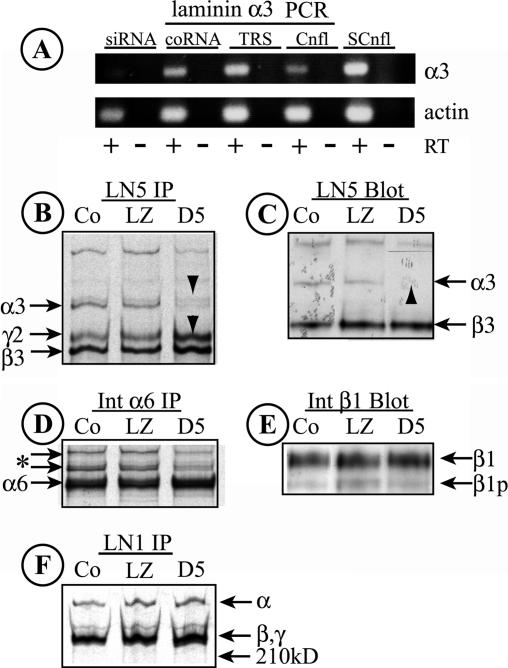
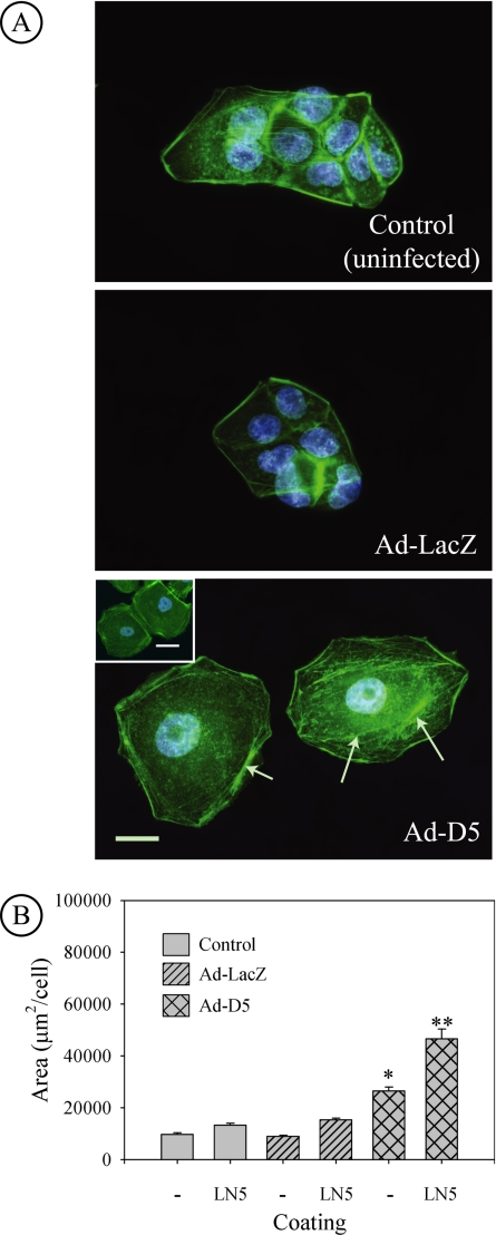
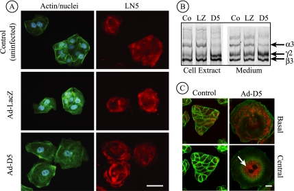
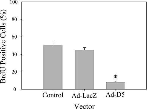
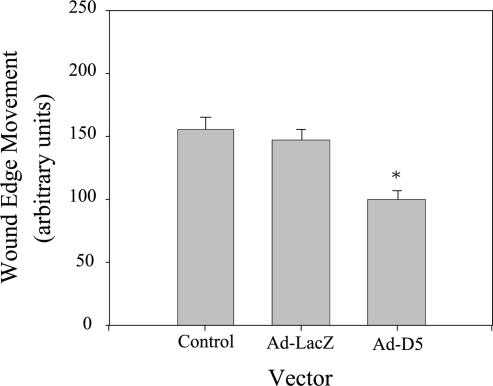
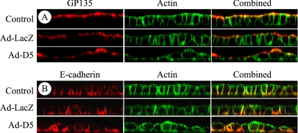
Renal tubular epithelial cells synthesize laminin (LN)5 during regeneration of the epithelium after ischemic injury. LN5 is a truncated laminin isoform of particular importance in the epidermis, but it is also constitutively expressed in a number of other epithelia. To investigate the role of LN5 in morphogenesis of a simple renal epithelium, we examined the synthesis and function of LN5 in the spreading, proliferation, wound-edge migration, and apical-basal polarization of Madin-Darby canine kidney (MDCK) cells. MDCK cells synthesize LN5 only when subconfluent, and they degrade the existing LN5 matrix when confluent. Through the use of small-interfering RNA to knockdown the LN5 alpha3 subunit, we were able to demonstrate that LN5 is necessary for cell proliferation and efficient wound-edge migration, but not apical-basal polarization. Surprisingly, suppression of LN5 production caused cells to spread much more extensively than normal on uncoated surfaces, and exogenous keratinocyte LN5 was unable to rescue this phenotype. MDCK cells also synthesized laminin alpha5, a component of LN10, that independent studies suggest may form an assembled basal lamina important for polarization. Overall, our findings indicate that LN5 is likely to play an important role in regulating cell spreading, migration, and proliferation during reconstitution of a continuous epithelium.


Figures

Figure 1.
Figure 1.
LN5 is only expressed in MDCK cells at early times after plating. (A) Endogenous matrix proteins deposited by MDCK cells (8 × 10⁶ cells/10-cm2 culture dish) for the indicated times were immunoblotted with an antibody against the laminin β3 subunit. Immunoreactive material corresponding to deposited LN5 was detected beginning at 6 h plating, with increasing amounts afterward. An apparent β3 degradation product was also observed (*). (B) Immunodetection of endogenous matrix proteins deposited on the culture dish by MDCK cells 18 h, 4 d, or 7 d after plating. Under these conditions, LN5 deposition declines significantly after 4 d and is nearly absent after 7 d. As in A, a β3 degradation product is visible (*). (C) MDCK cells (7.5 × 10⁵ cells/35-mm dish) were plated on coverslips and fixed and stained with a polyclonal antibody against LN5 at either 18 h, 4 d, or 7 d after plating. Coverslips were observed by confocal fluorescence microscopy; only sections through the center of the cell are shown. After 18 h, LN5 staining (red) is apparent throughout the cytoplasm in a pattern resembling the endoplasmic reticulum. By 4 d, only a few cytoplasmic vesicles per cell are visible, with only isolated staining (arrows) by 7 d. Green, actin. Bar, 10 μm. (D) Pulse labeling and immunoprecipitation of LN5 in MDCK cells. MDCK cells (1 × 10⁶ cells/35-mm dish) were pulse labeled for 30 min at the indicated times after plating with 50 μCi of [35S]Met/Cys. After labeling, the cells were extracted with RIPA buffer, the extracts were immunoprecipitated with polyclonal anti-LN5, and the immunoprecipitates were analyzed by SDS-gel electrophoresis and fluorography. After 18 h plating, all three LN5 subunits are detected, whereas little α3 or γ2 is detected at later times.
Figure 2.
Figure 2.
MDCK cells synthesize the laminin α5 subunit. (A) PCR products corresponding to laminin α5 and β-actin were detected in RNA from MDCK cells by RT-PCR using primers specific to human laminin α5 and canine β-actin. Lanes 1 and 2, laminin α5 primers; lane 3, actin primers. (B) The PCR product shown in A was sequenced and the sequence compared with that of human and mouse laminin α5. The canine sequence is 85% identical to the human sequence and 81% identical to the mouse sequence. (C) MDCK cells (7.5 × 10⁶/35-mm dish) were plated for 42 h and then fixed and stained for either LN5 or putative LN10, the latter using a polyclonal antibody against LN1. Confocal sections from either the extreme base of the cells (top) or through the center of cells (bottom) are illustrated. In the case of LN5, significant amounts of deposited LN5 are visible at the base, but the cytoplasm has only limited amounts of perinuclear staining. For LN10, both the base and cytoplasmic compartments are strongly stained, indicating not only deposition of the protein but also continued synthesis. Bar, 20 μm.
Figure 3.
Figure 3.
MDCK cells use β1 integrins and α6β4 integrin to adhere to LN5. A single-cell suspension of MDCK cells was plated in wells of a 96-well plate coated with either bovine serum albumin (BSA) (−control), collagen I, or LN5 in the presence or absence of function-blocking antibodies against β1 integrin (AIIB2) or α6 integrin (GoH3). After 90-min incubation at 37°C, unattached cells were washed away, and numbers of attached cells were estimated after fixation and staining by measuring absorbance of solubilized stain. Almost no cells attach to BSA-coated wells, whereas nearly all cells attach to wells coated with either collagen I or LN5. Anti-β1 integrin abolishes all adhesion to collagen I, but by itself has no effect on adhesion to LN5. Anti-α6 integrin reduces cell adhesion to LN5 somewhat, but is much more effective in combination with anti-β1 integrin. *p < 0.05 and **p < 0.001 relative to no antibody LN5 control.
Figure 4.
Figure 4.
MDCK cells spread extensively on LN5 using a β1 integrin. (A) MDCK cells were plated on coverslips coated with collagen I (i) or LN5 (ii–iv) for 2 h at 37°C in the presence or absence of function-blocking anti-β1 integrin (AIIB2; iii) or α6 integrin (GoH3; iv). At the end of the incubation, the cells were fixed and stained with fluorescent phalloidin and viewed by Nomarski DIC microscopy (i–iv) or fluorescence microscopy (ii; inset). On collagen, cells spread asymmetrically and extend narrow lamellipodia (i); on LN5, cells spread more extensively and symmetrically than on collagen (ii; arrowheads mark cell edges). By fluorescence, the spread cells on LN5 display thick bundles of filamentous actin at the cell periphery (ii; inset). In the presence of anti-β1 integrin, cells attach to LN5 but do not spread, and blebs are present at the cell peripheries (arrowheads; iii). In the presence of anti-α6 integrin, the cells on LN5 spread but not as extensively or uniformly as in the absence of antibody (iv). Bar, 10 μm. (B) MDCK cells were plated at subconfluent density for 24 h at 37°C on uncoated glass coverslips or coverslips coated with either collagen I or LN5. At the end of the incubation, the cells were fixed and stained with fluorescent phalloidin and DAPI. The area occupied by spread cells was then measured using MetaMorph. Cells plated on LN5 spread significantly more than those plated on either glass or collagen I. *p < 0.001 relative to uncoated control and collagen coating.
Figure 5.
Figure 5.
Knockdown of laminin α3 with siRNA. (A) RNA was harvested from MDCK cells transfected for 2 d with either a synthetic RNA duplex specific for a target sequence in canine laminin α3 (siRNA), a control duplex not representing any known gene (coRNA), or transfection reagent alone (TRS). RNA was also harvested from confluent (Cnfl) or subconfluent (SCnfl) MDCK cell cultures. Laminin α3 and actin transcripts were detected by RT-PCR. RT, reverse transcriptase added to the reaction. Note the absence of a laminin α3 band with transfected siRNA. (B) LN5 was immunoprecipitated from detergent extracts of metabolically labeled control MDCK cells (Co) or cells infected with siRNA-expressing adenovirus (D5) or a control virus (LZ). In both Co and LZ samples, all three LN5 bands are visible; in the D5 siRNA sample, little laminin α3 can be seen, whereas the levels of laminin β3 and γ2 chains are increased (arrowheads). (C) Extracts from control (Co), Ad-LacZ–infected, and Ad-D5–infected MDCK cells were immunoblotted with anti-LN5. Laminin α3, which is weakly detected by the antibody, is absent in cells expressing siRNA (arrowhead). The laminin γ2 chain is not detected by the antibody. (D and F) The labeled cell extracts used to immunoprecipitate LN5 in B were sequentially immunoprecipitated with anti-α6 integrin (D) and anti-LN1 (F). No significant differences between the samples are evident, suggesting that siRNA-mediated knockdown of laminin α3 is specific. The two bands in D running more slowly than α6 (* and arrows) are unidentified but may correspond to forms of integrin β4. In F, the higher molecular weight band is likely laminin α5 and the lower doublet β1 and γ1. (E) The immunoblot shown in C was stripped and reprobed with anti-β1 integrin. No significant differences between samples can be seen. β1, mature β1 integrin; β1p, precursor β1.
Figure 6.
Figure 6.
Knockdown of laminin α3 leads to increased spreading of MDCK cells. (A) Uninfected MDCK cells and cells infected with control Ad-LacZ virus or siRNA-expressing Ad-D5 virus were replated on uncoated glass coverslips overnight and then fixed and stained with fluorescent phalloidin (green) and DAPI (blue) to visualize the actin cytoskeleton and nuclei, respectively. Micrographs show that cells infected with Ad-D5 spread much more than either control or Ad-LacZ–infected cells and are so flat that the nucleus is distorted and seems larger. Bundles of actin filaments and extensive actin stress fibers are also apparent (arrows). Bar, 20 μm. Inset in Ad-D5 panel, formation of cell–cell contacts does not suppress extensive cell spreading. Bar, 20 μm. (B) Control MDCK cells or cells infected with Ad-LacZ or Ad-D5 were replated on uncoated glass coverslips (−) or coverslips coated with purified human LN5 (LN5). Fixed cells were stained with fluorescence phalloidin and DAPI, and spread area/cell was measured from digital micrographs. Cells expressing siRNA targeted against laminin α3 spread much more than either control or Ad-LacZ–infected cells. Spreading of all cells is stimulated further by plating on LN5-coated coverslips. *p < 0.05 relative to uncoated control and Ad-LacZ. **p < 0.05 relative to LN5-coated control and Ad-LacZ replated on LN5. Ad-D5 on uncoated glass (*) compared with Ad-D5 on LN5-coated glass (**p < 0.001 by t test).
Figure 7.
Figure 7.
Laminin β3 and γ2 are secreted after siRNA-mediated knockdown of laminin α3. (A) Control MDCK cells or cells infected for 18 h with Ad-LacZ or Ad-D5 were replated on uncoated glass coverslips for 24 h and stained with antibodies against LN5 (red), fluorescent phalloidin (green), and DAPI (blue), and viewed by conventional immunofluorescence microscopy. Anti-LN5 antibody staining is visible at the base in all three cases, although the staining pattern under cells expressing siRNA (Ad-D5) exhibits a characteristic “rose petal” pattern. Bar, 20 μm. (B) Control MDCK cells or cells infected with Ad-LacZ or Ad-D5 were metabolically labeled overnight. Detergent cell extracts and culture medium were immunoprecipitated with anti-LN5, and precipitated polypeptides were visualized on SDS-gels by fluorography. In extracts from Ad-D5–infected cells, the laminin α3 band is absent and laminin β3 and γ2 are increased in intensity (D5 lane). In the medium, laminin α3 is detectable but diminished, and the ratio of the β3 and γ2 bands to α3 is higher than controls, suggesting secretion of β3 and γ2 uncomplexed to α3 (compare D5 with Co and LZ). (C) Control MDCK cells or cells infected for 42 h with Ad-D5 were replated on uncoated glass coverslips for 24 h, stained with antibodies against LN5 (red) and fluorescent phalloidin (green), and viewed by confocal fluorescence microscopy. Individual optical sections taken at either the base of the cells or through the cell center are shown. Cells infected with Ad-D5 are exceedingly spread and still exhibit some staining with anti-LN5 at the base of the cell. Inside the same cells, anti-LN5 staining is intense in what seems to be the endoplasmic reticulum (arrow). Control (uninfected) cells seem normal with some anti-LN5 staining both within the cell and at the cell base. Ad-LacZ–infected cells seemed identical to uninfected controls (our unpublished data). Bar, 10 μm.
Figure 8.
Figure 8.
Knockdown of laminin α3 blocks proliferation. Control MDCK cells or cells infected for 18 h with Ad-LacZ or Ad-D5 were replated on uncoated glass coverslips for 24 h, incubated with BrdU for 6 h, stained with antibodies against BrdU and DAPI (blue), and viewed by conventional immunofluorescence microscopy. Proliferation was measured by counting total and BrdU-positive nuclei. *p < 0.05.
Figure 9.
Figure 9.
Knockdown of laminin α3 slows wound-edge migration. Control MDCK cells or cells infected for 18 h with Ad-LacZ or Ad-D5 were replated at confluent density on gridded, uncoated glass coverslips for 24 h, and wounded by scraping. Wound-edge migration relative to the time of wounding was measured on micrographs after 24 h. Note that wound-edge movement occurred under all three conditions but was significantly reduced in cells expressing siRNA (Ad-D5). *p < 0.05.
Figure 10.
Figure 10.
Knockdown of laminin α3 affects epithelial morphogenesis but not apical–basal polarization. Control MDCK cells or cells infected for 18 h with Ad-LacZ or Ad-D5 were replated at confluent density on uncoated Transwell supports for 24 h and stained with antibodies against either the apical antigen gp135 (A; red) or the basolateral protein E-cadherin (B; red) and fluorescent phalloidin (green). The stained samples were imaged as orthogonal Z-sections by confocal fluorescence microscopy. Note that the epithelium formed by Ad-D5–infected cells is heteromorphic with cells of different sizes and shapes, but it is as polarized relative to both apical and basolateral proteins as control and Ad-LacZ–infected cultures. Cell height in all samples is ∼10–15 μm.

Similar articles

Cited by

References

    1. Aumailley M., El Khal A., Knoss N., Tunggal L. Laminin 5 processing and its integration into the ECM. Matrix Biol. 2003;22:49–54. - PubMed
    1. Boll W., Partin J. S., Katz A. I., Caplan M. J., Jamieson J. D. Distinct pathways for basolateral targeting of membrane and secretory proteins in polarized epithelial cells. Proc. Natl. Acad. Sci. USA. 1991;88:8592–8596. - PMC - PubMed
    1. Caplan M. J., Stow J. L., Newman A. P., Madri J., Anderson H. C., Farquhar M. G., Palade G. E., Jamieson J. D. Dependence on pH of polarized sorting of secreted proteins. Nature. 1987;329:632–635. - PubMed
    1. Carter W. G., Ryan M. C., Gahr P. J. Epiligrin, a new cell adhesion ligand for integrin α3β1 in epithelial basement membranes. Cell. 1991;65:599–610. - PubMed
    1. Cheng Y. S., Champliaud M. F., Burgeson R. E., Marinkovich M. P., Yurchenco P. D. Self-assembly of laminin isoforms. J. Biol. Chem. 1997;272:31525–31532. - PubMed

Publication types

LinkOut - more resources