Comparative Study
. 2006 Jun 14;26(24):6627-36.
doi: 10.1523/JNEUROSCI.0149-06.2006.

Subventricular zone-derived neuroblasts migrate and differentiate into mature neurons in the post-stroke adult striatum

Affiliations
Comparative Study

Subventricular zone-derived neuroblasts migrate and differentiate into mature neurons in the post-stroke adult striatum

Toru Yamashita et al. J Neurosci. .

Abstract

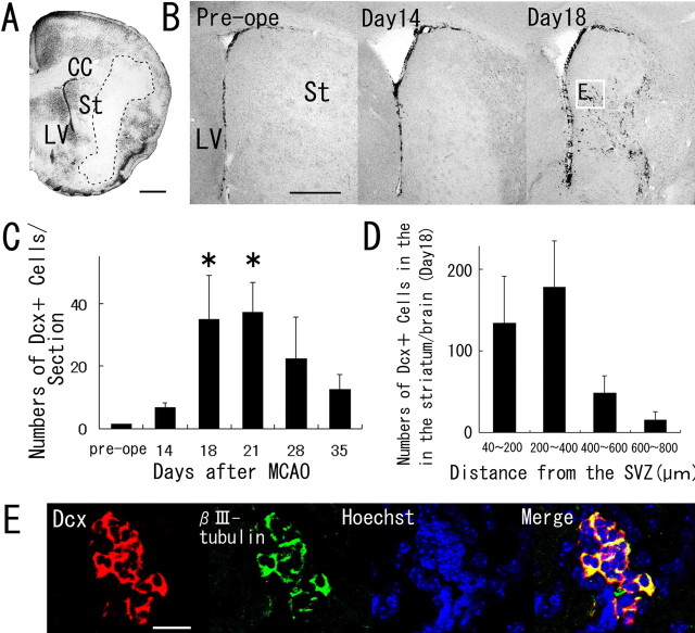
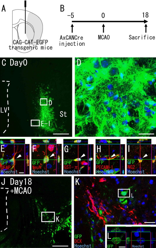
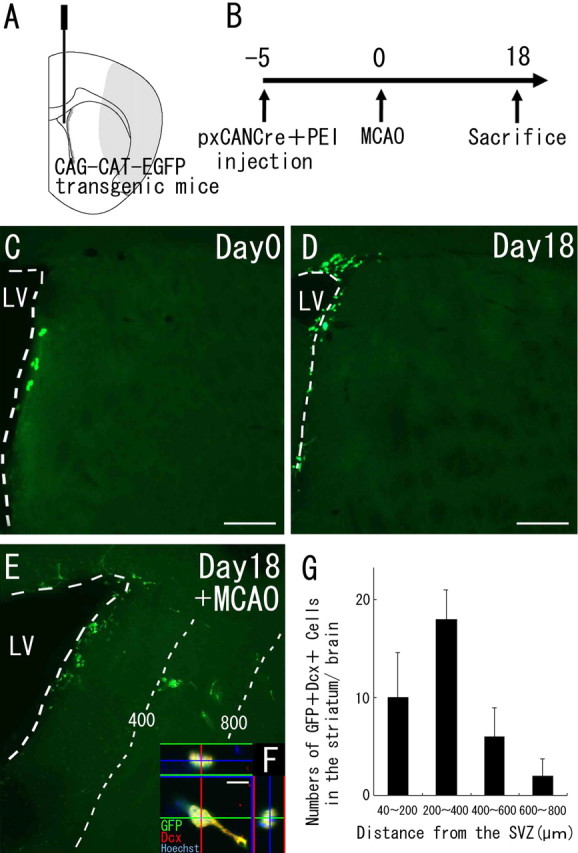
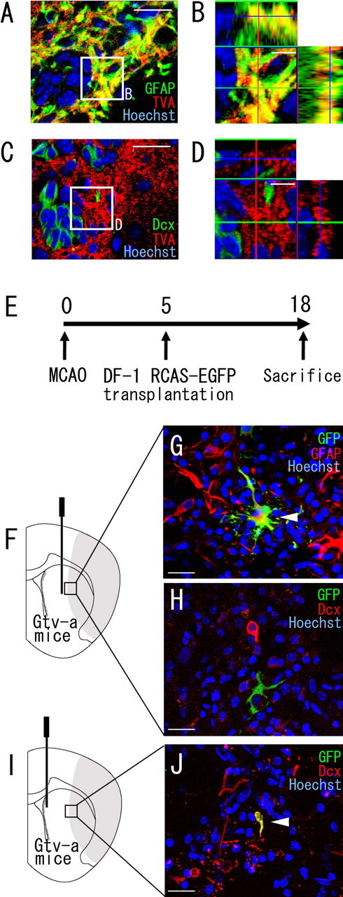
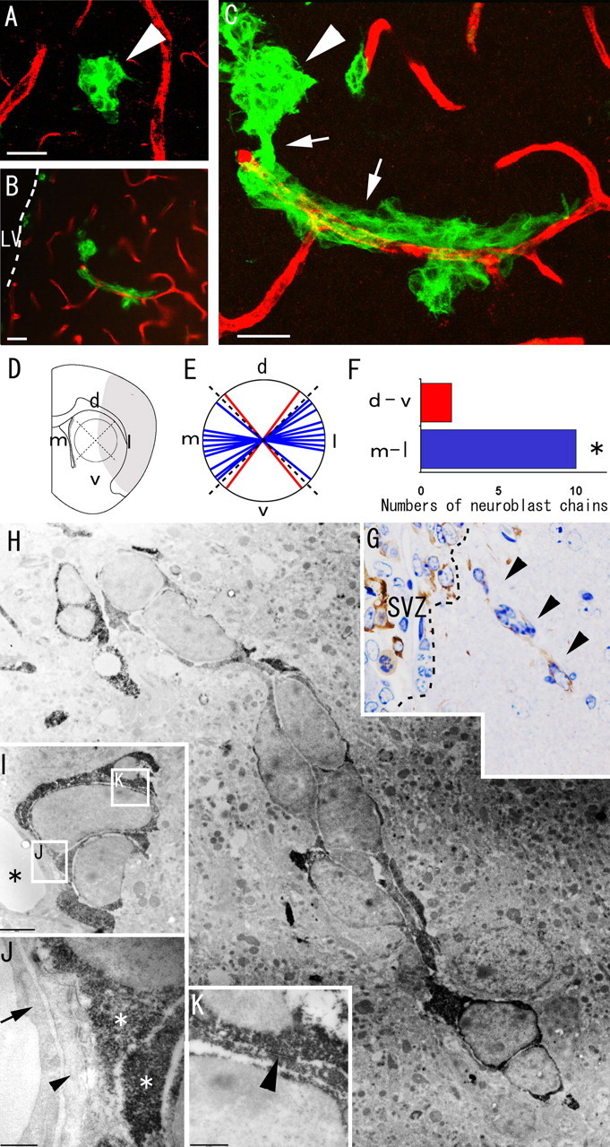
Recent studies have revealed that the adult mammalian brain has the capacity to regenerate some neurons after various insults. However, the precise mechanism of insult-induced neurogenesis has not been demonstrated. In the normal brain, GFAP-expressing cells in the subventricular zone (SVZ) of the lateral ventricles include a neurogenic cell population that gives rise to olfactory bulb neurons only. Herein, we report evidence that, after a stroke, these cells are capable of producing new neurons outside the olfactory bulbs. SVZ GFAP-expressing cells labeled by a cell-type-specific viral infection method were found to generate neuroblasts that migrated toward the injured striatum after middle cerebral artery occlusion. These neuroblasts in the striatum formed elongated chain-like cell aggregates similar to those in the normal SVZ, and these chains were observed to be closely associated with thin astrocytic processes and blood vessels. Finally, long-term tracing of the green fluorescent-labeled cells with a Cre-loxP system revealed that the SVZ-derived neuroblasts differentiated into mature neurons in the striatum, in which they expressed neuronal-specific nuclear protein and formed synapses with neighboring striatal cells. These results highlight the role of the SVZ in neuronal regeneration after a stroke and its potential as an important therapeutic target for various neurological disorders.


Figures

Figure 1.
Figure 1.
Neuroblasts transiently appearing in the striatum after cerebral ischemia. A, A coronal brain section, obtained 14 d after ischemia induction and stained with cresyl violet. Dotted lines indicate the border between the intact and infarcted areas. CC, Corpus callosum; St, striatum; LV, lateral ventricle. Scale bar, 500 μm. B, Anti-Dcx staining of coronal brain sections obtained preoperatively (Pre-ope) and at 14 and 18 d after MCAO. The boxed area in B indicates the field shown in E. Scale bar, 500 μm. C, Temporal profile of Dcx-positive cells in the striata of MCAO mice (n = 3 at each time point). The number of Dcx-positive cells at 18 and 21 d after ischemia induction was significantly greater than the preoperative number. Values are means ± SEM. ∗p < 0.01 versus preoperative value (ANOVA, Bonferroni’s correction). D, Numbers of Dcx-positive cells in four regions at different distances from the SVZ of brains (n = 3) 18 d after MCAO. E, Double immunohistochemistry of the striatum with anti-Dcx antibody (red) and anti-βIII-tubulin antibody (green) 18 d after ischemia induction. Another neuroblast marker, βIII-tubulin, was coexpressed by 98% of the Dcx-positive cells (n = 1108 cells) in the striatum. Nuclear staining with Hoechst 33258 is shown in blue. Scale bar, 50 μm.
Figure 2.
Figure 2.
Striatal cells do not generate neuroblasts. A, Diagram showing the injection site. AxCANCre, an adenoviral vector encoding Cre recombinase under control of the CAG promoter, was injected into the striata of CAG-CAT-EGFP transgenic mice, which carry a floxed EGFP gene under control of the CAG promoter. B, AxCANCre was injected 5 d before MCAO, and the animals were killed 18 d after MCAO. C–I, Expression of GFP in the striatum on day 0 (5 d after injection). C, Large numbers of GFP-positive cells (green) are present in the striatum, although there are none in the SVZ. Scale bar, 500 μm. LV, Lateral ventricle; St, striatum. D–I, High-power confocal images of the injection site indicated by the boxed areas in C. D, All Hoechst 33258-positive cells (blue) express detectable GFP (green) in the vicinity of the injection site. Scale bar, 20 μm. E–I, Double stainings with cell-type-specific markers reveal that the GFP-encoding adenovirus-infected cells express GFAP (E), NeuN (F) GST-π (G), PECAM-1 (H), and NG2 (I). Scale bar, 10 μm. Arrowheads point to GFP-positive cells colabeled with the indicated marker. J, Distribution of GFP-positive cells (green) in the striatum 18 d after MCAO. Scale bar, 500 μm. K, High-power confocal image of the injection site indicated by the boxed area in J. All GFP-labeled cells (n = 695) observed in three MCAO mice are negative for Dcx. Scale bar, 20 μm. L, Orthogonal view of a GFP-positive cell indicated by the boxed area in K. Among all of the GFP-positive cells analyzed (n = 62), each cell had an intact nucleus. Scale bar, 10 μm.
Figure 3.
Figure 3.
Neuroblasts migrate from the SVZ into the striatum after cerebral ischemia. A, Diagram showing the injection site. The Cre-encoding pxCANCre plasmid was injected into the lateral ventricles of CAG-CAT-EGFP transgenic mice, which carry the floxed EGFP gene under control of the CAG promoter. B, pxCANCre plasmid plus PEI was injected into the lateral ventricle 5 d before MCAO, and the animals were killed 18 d after MCAO. C, D, In the normal brain (n = 4 at each time point), GFP-positive cells (green) are observed only in the SVZ at 5 and 23 d after the injection (corresponding to days 0 and 18, respectively, in B). Scale bars, 200 μm. E, In the ischemic brain (n = 4), many SVZ-derived GFP-positive cells (green) are observed in the peri-infarct striatum 23 d after the injection (corresponding to day 18 in B). The numbers and thin dotted lines indicate distance (in micrometers) from the SVZ. F, High-power confocal image of a GFP/Dcx-expressing cell exhibiting morphology typical of migrating neuroblasts. Thirty-seven percent of the GFP-positive cells (n = 45) in the striatum expressed Dcx. Scale bar, 10 μm. G, Numbers of GFP/Dcx double-positive cells in four regions at different distances from the SVZ 18 d after MCAO. This distribution is very similar to those of GFP-positive cells (Fig. 1D). No SVZ-derived GFP/Dcx double-immunopositive cells were observed in the striatum of the non-ischemic brain.
Figure 4.
Figure 4.
SVZ GFAP-expressing cells are a source of the neuroblasts that migrate into the striatum after cerebral ischemia. A–D, Double immunofluorescence of an SVZ section stained with anti-TVA antibody (red), in combination with anti-GFAP antibody (green) (A, B) or with anti-Dcx antibody (green) (C, D) 8 d after induction of ischemia (Gtv-a transgenic mice). B and D show high-power views of the field indicated by the boxed areas in A and C, respectively. Note that all of the TVA-expressing cells contain detectable signals of GFAP but not Dcx. B, D, 10 μm. E, DF-1 cells transfected with the RCAS-EGFP plasmid were transplanted into the striatum (F–H) or SVZ (I, J) 5 d after MCAO, and the animals were killed 18 d after MCAO. F–H, In the brains (n = 16) in which RCAS-EGFP-producing DF-1 cells were transplanted into the striatum (F), GFP/GFAP double-positive cells were frequently observed (G). However, all GFP+ cells examined (n = 498) were negative for Dcx (H). Scale bar, 20 μm. I, J, All brains (n = 3) in which RCAS-EGFP-producing DF-1 cells were transplanted into the SVZ (I) contained GFP/Dcx double-positive cells (5 cells per 13 GFP-positive cells in total) in the peri-infarct region of the striatum at 18 d (J). Scale bar, 20 μm.
Figure 5.
Figure 5.
Migrating neuroblasts formed elongated aggregates associated with blood vessels in the ischemic striatum. A–C, Double immunohistochemistry of the striatum with anti-PECAM-1 antibody (red) and anti-Dcx antibody (green) 18 d after ischemia induction. A, Spherical clusters of Dcx-positive neuroblasts (n = 6) are at some distance from the PECAM-1-positive vascular endothelial cells. B, C, Chains of neuroblasts (n = 12) wind around the vascular endothelial cells. Note that this chain appears to extend toward the injured region (on the right, in this figure) and to be connected with a spherical cluster of neuroblasts that is not associated with blood vessels. See also supplemental movie 1 (available at www.jneurosci.org as supplemental material). Arrowheads, Spherical cluster of neuroblasts; arrows, elongated chain-like aggregate of neuroblasts. LV, Lateral ventricle. Scale bars, 20 μm. D, The chains were classified on the basis of their dorsoventral (d-v) and mediolateral (m-l) orientations. d, Dorsal; v, ventral; m, medial; l, lateral. E, F, The number of mediolaterally oriented chain-like neuroblasts (blue) was significantly larger than the number of dorsoventrally oriented neuroblasts (red). ∗p < 0.05 (χ2 test). G, Dcx-stained semithin section of the striatum. The ectopic chain of neuroblasts in the electron micrograph in H is marked by arrowheads. H–K, Electron microscopic view of neuroblasts migrating within the striatum. H, Each Dcx-positive cell contains abundant lax chromatin, two to four small nucleoli, and a smooth scant cytoplasm, similar to neuroblasts in the SVZ. I, Dcx-positive neuroblasts are located close to blood vessels. Asterisks, Blood vessels. The boxed areas in I indicate the fields shown in J and K. Scale bar, 2 μm. J, Dcx-positive neuroblasts are adjacent to the thin processes of astrocytes (arrowhead) associated with endothelial cells (arrow) of blood vessels. Asterisks, Cytoplasm of Dcx-positive neuroblasts. Scale bar, 500 nm. K, A zonula-adherens-like contact (arrowhead) is observed between two Dcx-positive neuroblasts. Scale bar, 500 nm.
Figure 6.
Figure 6.
SVZ-derived neuroblasts differentiate into mature neurons and form synapses in the striatum by 90 d after MCAO. A, Diagram showing the injection site. B, The pxCANCre plasmid was injected into the lateral ventricle 5 d before MCAO, and the animals were killed 90 d after MCAO. C, Confocal three-dimensional reconstruction image of a GFP/NeuN double-positive cell. At 90 d after MCAO, 29% of the SVZ-derived GFP-positive cells (n = 13) in the striatum expressed NeuN, a specific marker for mature neurons. Scale bar, 20 μm. D, A GFP-positive cell exhibiting neuronal morphology. Scale bar, 20 μm. E, An electron micrograph showing a GFP-positive axon (asterisk) containing presynaptic vesicles. Scale bar, 0.5 μm. F, High-power magnification view of the region indicated by the boxed area in E. The postsynaptic density is indicated by arrowheads.

References

    1. Altman J (1969). Autoradiographic and histological studies of postnatal neurogenesis. IV. Cell proliferation and migration in the anterior forebrain, with special reference to persisting neurogenesis in the olfactory bulb. J Comp Neurol 137:433–457. - PubMed
    1. Alvarez-Buylla A, Garcia-Verdugo JM (2002). Neurogenesis in adult subventricular zone. J Neurosci 22:629–634. - PMC - PubMed
    1. Araki T, Shibata M, Takano R, Hisahara S, Imamura S, Fukuuchi Y, Saruta T, Okano H, Miura M (2000). Conditional expression of anti-apoptotic protein p35 by Cre-mediated DNA recombination in cardiomyocytes from loxP-p35-transgenic mice. Cell Death Differ 7:485–492. - PubMed
    1. Arvidsson A, Kokaia Z, Lindvall O (2001). N-methyl-d-aspartate receptor-mediated increase of neurogenesis in adult rat dentate gyrus following stroke. Eur J Neurosci 14:10–18. - PubMed
    1. Arvidsson A, Collin T, Kirik D, Kokaia Z, Lindvall O (2002). Neuronal replacement from endogenous precursors in the adult brain after stroke. Nat Med 8:963–970. - PubMed

Publication types

MeSH terms