

Case Reports
Diffusion-weighted MR imaging in early diagnosis and prognosis of hypoglycemia
Affiliations
- PMID: 16775268
- PMCID: PMC8133946
Item in Clipboard
Case Reports
Diffusion-weighted MR imaging in early diagnosis and prognosis of hypoglycemia
AJNR Am J Neuroradiol.
2006 Jun-Jul.
Abstract
We describe 2 cases of diffusion-weighted (DW) MR imaging in hypoglycemic coma. One patient, with diffuse cortical lesions, had a poor outcome, but the other, with transient white matter abnormalities, made a complete recovery. The distinctive patterns of DW MR imaging abnormalities in hypoglycemic patients should be recognized and may be a predictor of clinical outcome.
Figures
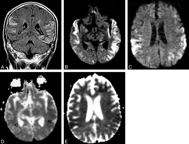
Case 1, a 65-year-old man in a diabetic coma with seizures. A, Fast spin-echo fluid attenuated inversion recovery (9000 milliseconds/110 milliseconds effective/2200 milliseconds [TR/TE/TI]) MR image shows bilateral hyperintensity of the cortex over the temporal and occipital lobes. B and C, Diffusion-weighted (10000/105, b value 1000 seconds/mm2) MR images showing corresponding hyperintensity in the cortex. D and E, ADC maps at the same levels as B and C show decreased ADC in these lesions (618 × 10−3 mm2/s) compared with normal white matter (819 × 10−3 mm2/s).
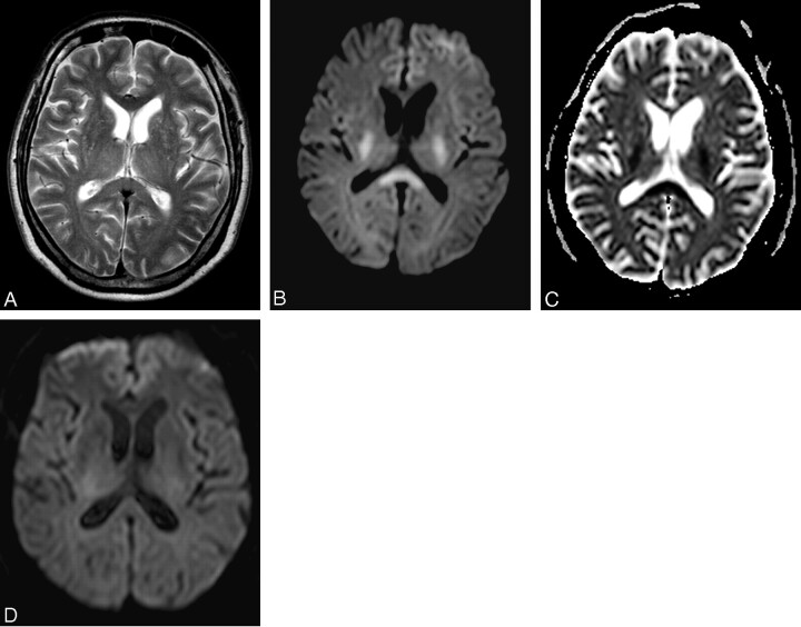
Case 2, a 69-year-old diabetic man with atrial fibrillation who suddenly became unresponsive. A, T2-weighted (3000/80 effective) MR image shows subtle increased intensity in the splenium of the corpus callosum (compared with the genu), posterior limbs of the internal capsules, and thalami bilaterally. B, Corresponding diffusion-weighted (4200/95, b value 1000 seconds/mm2) MR image shows bilaterally symmetrical hyperintensities in the posterior limbs of the internal capsules and the splenium of the corpus callosum. C, ADC map corresponding to B shows decreased ADC of the splenium of the corpus callosum (485 × 10−3 mm2/s) compared with the genu (890 × 10−3 mm2/s) D, DW MR image 12 hours later shows complete normalization of previously noted lesions.
References
-
- Lockwood AH. Toxic and metabolic encephalopathies. In: Bradley WG, Daroff RB, Fenichal GM, et al, eds. Neurology in clinical practice. 3rd ed. London: Butterworth-Heinemann;2000. :1485
-
- Boeve BF, Bell DG, Noseworthy JH. Bilateral temporal lobe MRI changes in uncomplicated hypoglycemic coma. Can J Neurol Sci 1995;22:56–58 - PubMed
-
- Fujioka M, Okuchi K, Hiramatsu KI, et al. Specific changes in human brain after hypoglycemic injury. Stroke 1997;28:584–87 - PubMed
-
- Finelli PF. Diffusion-weighted MR in hypoglycemic coma. Neurology 2001;57:933–35 - PubMed
-
- Aoki T, Sato T, Hasegawa K, et al. Reversible hyperintensity lesion on diffusion-weighted MRI in hypoglycemic coma. Neurology 2004;63:392–93 - PubMed
Publication types
MeSH terms
LinkOut - more resources
Full Text Sources
Medical