

Case Reports
Basilar and bilateral carotid dolichoectasia with spontaneous dissection of C2 segment of the internal carotid artery
Affiliations
- PMID: 16775273
- PMCID: PMC8133952
Item in Clipboard
Case Reports
Basilar and bilateral carotid dolichoectasia with spontaneous dissection of C2 segment of the internal carotid artery
AJNR Am J Neuroradiol.
2006 Jun-Jul.
Abstract
We describe a previously unreported case of cranial arterial dolichoectasia associated with spontaneous dissection of the petrous (C2) segment of the internal carotid artery (ICA) with 2 patent lumena. Dolichoectasia of the cranial arteries and different types of double lumen of ICA are discussed. A review of previously reported cases is included.
Figures
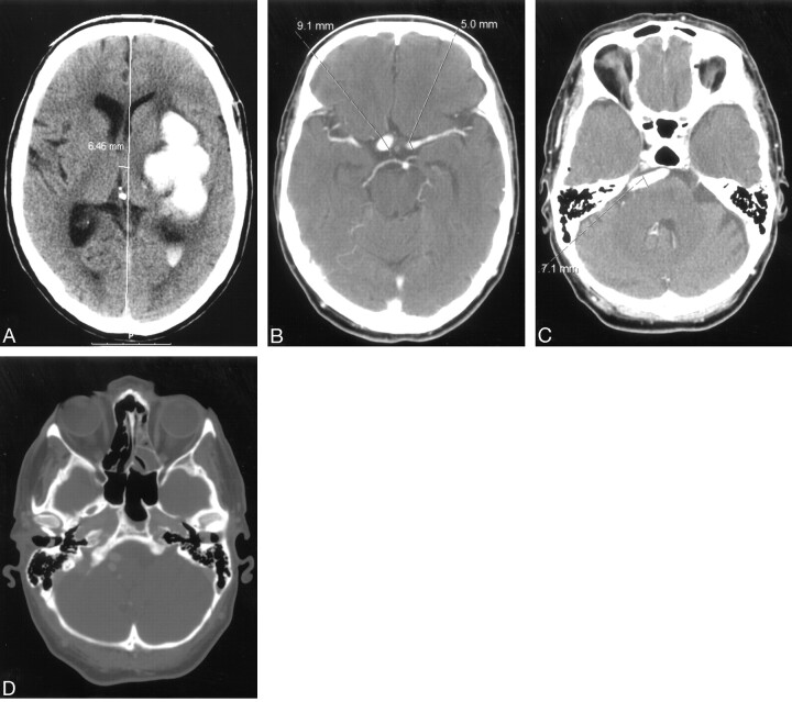
A, nonenhanced computerized tomography showing left-sided ganglionic bleeding with penetration of the blood into the left lateral ventricle. B and C, contrast-enhanced computerized tomography (CECT) showing supraclionoid segments of both internal carotid arteries and tortuous and dilated basilar artery. D, CECT, bone window shows symmetrical and normally shaped carotid channels within hyperaerated petrous bones.
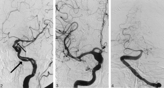
Angiogram of the left internal carotid artery, left anterior oblique projection showing double lumen of C2 segment of the internal carotid artery. Note the smooth contours of slightly dilated vascular channels (arrow) and fusiform dilation of the supraclinoid segment of the internal carotid artery (small arrow).
Angiogram of the right internal carotid artery, anteroposterior (AP) projection, shows symmetric fusiform dilation of the supraclinoid segment of the right internal carotid artery.
Angiogram of the basilar artery, AP projection, shows moderately dilated distal segment of the left vertebral artery as well as dilated and extremely tortuous basilar artery.
References
-
- Courville CB. Arteriosclerotic aneurysms of the circle of Willis: some notes of their morphology and pathogenesis. Bull Los Angeles Neurol Soc 1962;27:1–13 - PubMed
-
- Ince B, Petty GW, Brown RD, et al. Dolichoectasia of the intracranial arteries in patients with first ischemic stroke: a population-based study. Neurology 1998;50:1694–97 - PubMed
-
- Sacks JG, Lindenburg R. Dolicho-ectatic intracranial arteries: symptomatology and pathogenesis of arterial elongation and distension. Johns Hopkins Med J 1969;125:95–106 - PubMed
-
- Moseley IF, Holland IM. Ectasia of the basilar artery, the breadth of the clinical spectrum, and the diagnostic value of computed tomography. Neuroradiology 1979;18:83–91 - PubMed
Publication types
MeSH terms
LinkOut - more resources
Full Text Sources
Medical
Miscellaneous