

Silent ischemia after neuroprotected percutaneous carotid stenting: a diffusion-weighted MRI study
- PMID: 16775293
- PMCID: PMC8133917
Silent ischemia after neuroprotected percutaneous carotid stenting: a diffusion-weighted MRI study
Abstract
Background and purpose: To assess by diffusion-weighted MR imaging (DWI) the efficacy of cerebral protection devices in avoiding embolization and new ischemic lesions in patients with severe internal carotid artery (ICA) stenosis undergoing carotid artery stent placement (CAS).
Methods: One hundred sixty-two CASs in the extracranial ICA were performed with the use of distal filters. Mean age of the patients was 68.5 years (range, 33-86) and 122 patients (75.3%) were symptomatic. MR imaging was performed in all patients during the 3-day period before CAS, and DWI was obtained within 24 hours after the procedure. Ninety-five patients (58.6%) were monitored by transcranial Doppler ultrasonography for microemboli detection in the territory of the middle cerebral artery (MCA), ipsilateral to the vessel being treated.
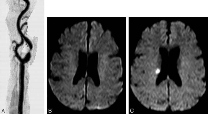
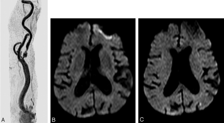
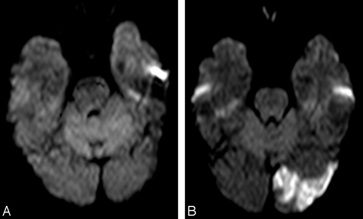
Results: Twenty-eight patients (17.3%) showed 58 new ischemic foci in DWI, and 13 patients (46.4%) had multiple foci. Location of new lesions was mainly in the vascular territory supplied by the treated vessel (19 patients; 67.9%), but also in the contralateral MCA (1 patient; 3.6%), and the posterior fossa (4 patients; 14.3%). A significant relationship (P < .03) was found between occurrence of transient ischemic attack (TIA) and appearance of new lesions. Microembolic signals (MES) were detected in 88 patients (92.6%), with no relationship between number of MES and the appearance of new ischemic foci.
Conclusion: New ischemic foci were observed in 17.3% of the patients undergoing neuroprotected CAS. Appearance of new ischemic lesions were only significantly related to the occurrence of TIA but not to the number of MES registered or other variables. Despite the encouraging results, the incidence of new ischemic lesions should promote research for safer techniques and devices.
Figures


References
-
- Yadav JS, Wholey MH, Kuntz RE, et al. Protected carotid-artery stenting versus endarterectomy in high-risk patients. N Engl J Med 2004;351:1493–501 - PubMed
-
- Ohki T, Marin ML, Lyon RT. Ex vivo human carotid artery bifurcation stenting:correlation of lesion characteristics with embolic potential. J Vasc Surg 1998;27:463–71 - PubMed
-
- Manninen HI, Rasanen HT, Vanninen RL, et al. Stent placement versus percutaneous transluminal angioplasty of human carotid arteries in cadavers in situ: distal embolization and findings at intravascular US, MR imaging and histopathological analysis. Radiology 1999;212:483–92 - PubMed
-
- Jordan WD, Voellinger DC, Doblar DD, et al. Microemboli detected by transcranial Doppler monitoring in patients during carotid angioplasty versus carotid endarterectomy. Cardiovasc Surg 1999;7:33–38 - PubMed
-
- Theron JG, Payelle GG, Coskun O, et al. Carotid artery stenosis: Treatment with protected balloon angioplasty and stent placement. Radiology 1996;201:627–36 - PubMed
Publication types
MeSH terms
LinkOut - more resources
Full Text Sources
Miscellaneous