

Case Reports
Transvenous treatment of spontaneous dural carotid-cavernous fistulas using a combination of detachable coils and Onyx
Affiliations
- PMID: 16775294
- PMCID: PMC8133910
Item in Clipboard
Case Reports
Transvenous treatment of spontaneous dural carotid-cavernous fistulas using a combination of detachable coils and Onyx
AJNR Am J Neuroradiol.
2006 Jun-Jul.
Abstract
Three patients with spontaneous dural carotid-cavernous fistulas were treated by using a combination of detachable coils and Onyx liquid embolic agent. Cavernous sinus was accessed via the superior ophthalmic vein or inferior petrous sinus approach. In all cases, a complete angiographic closure of the fistulas was achieved with full recovery from neuro-ophthalmologic symptoms. This report suggests that the controlled and excellent penetration of Onyx is superb for blocking the intricate communication of dural carotid-cavernous fistulas.
Figures
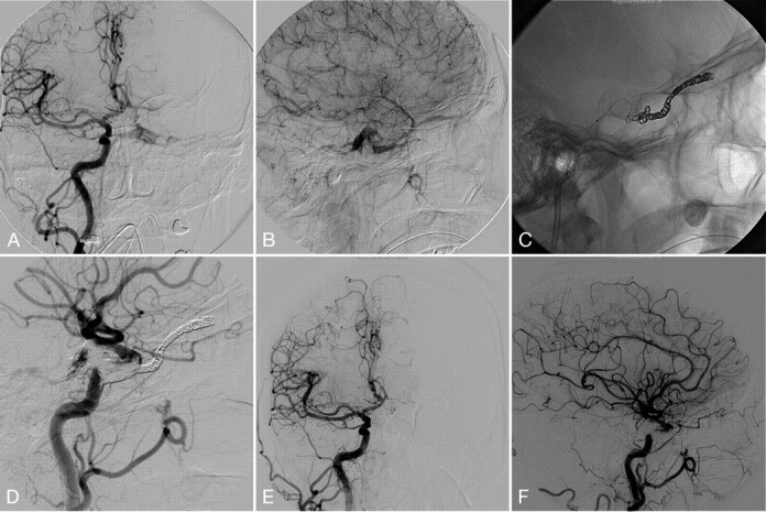
A, Pre-embolization right CCA. Frontal projection arterial phase shows the dural CCF with early venous drainage into the left cavernous sinus followed by the left IPS. B, Lateral projection of right CCA injection late capillary phase shows retrograde filling of abnormally dilated left SOV. C, Unsubtracted lateral projection image demonstrates transjugular-IPS venous access to the left SOV and deployed coils in the left SOV. D, Lateral view of the right CCA injection shows the repositioned microcatheter to the anterior compartment of the cavernous sinus and partial embolization with Onyx. E and F, Postembolization CCA angiogram. Frontal projection (E) and lateral projection (F) demonstrate complete obliteration of the fistula. Deployed coils in the left SOV and Onyx cast in the left cavernous and coronary sinuses block the fistula. There is no residual fistula.
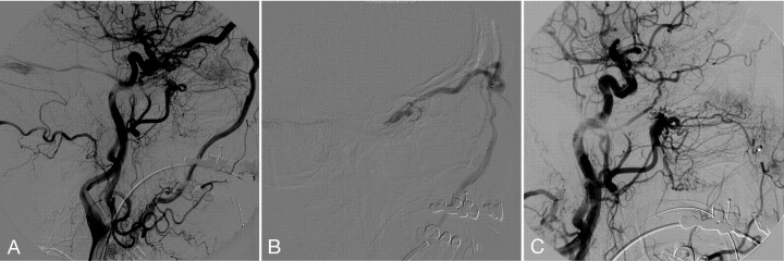
A, Left CCA angiogram lateral projection shows dural CCF fed by meningohypophyseal artery, accessory middle meningeal artery, and artery of foramen rotundum. The venous drainages are abnormally dilated left SOV and superior petrosal sinus. The IPS appeared occluded. B, Superselective venogram of the left SOV lateral view demonstrates multiple catheter support system with a 6F angled Envoy guiding catheter in the proximal left facial vein, a 4F Tracker 38 catheter in the left angular vein, and an Echelon 10 microcatheter. C, Postembolization with coils and Onyx angiogram of the left CCA shows complete occlusion of the fistula with no residual filling.
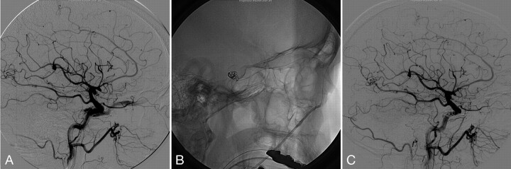
A, Right CCA injection lateral projection demonstrates dural CCF, with feeders from meningohypophyseal trunk with early venous drainage into the right SOV. The IPS is not visualized. No contribution of ECA was appreciated. B, Transexternal jugular-facial venous access is seen on the lateral projection with a 5F Glidecath XP in the right facial vein and a Prowler 14 microcatheter in the cavernous sinus/SOV junction, and deployed coils. C, Postprocedure right CCA angiogram lateral projection shows complete occlusion of the fistula with deployed coil mass and Onyx cast. A small piece of dislodged Onyx cast is also shown in the distal segment of right SOV.
References
-
- Murayama Y, Vinuela F, Ulhoa A, et al. Nonadhesive liquid embolic agent for cerebral arteriovenous malformations: preliminary histopathological studies in swine rete mirabile. Neurosurgery 1998;43:1164–75 - PubMed
-
- Jahan R, Murayama Y, Gobin YP, et al. Embolization of arteriovenous malformations with onyx: clinicopathological experience in 23 patients. Neurosurgery 2001;48:984–95; discussion 995–87 - PubMed
-
- Klisch J, Huppertz HJ, Spetzger U, et al. Transvenous treatment of carotid cavernous and dural arteriovenous fistulae: results for 31 patients and review of the literature. Neurosurgery 2003;53:836–56; discussion 856–37 - PubMed
Publication types
MeSH terms
Substances
LinkOut - more resources
Full Text Sources