Comparative Study
. 2006 Aug;12(8):1505-13.
doi: 10.1261/rna.2321606. Epub 2006 Jun 14.

Ribosomal protein S1 promotes transcriptional cycling

Affiliations
Comparative Study

Ribosomal protein S1 promotes transcriptional cycling

Maxim V Sukhodolets et al. RNA. 2006 Aug.

Abstract

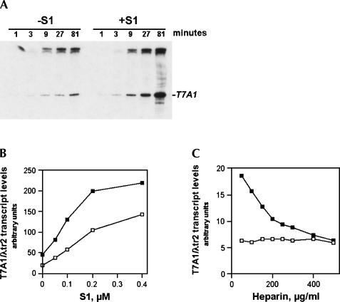
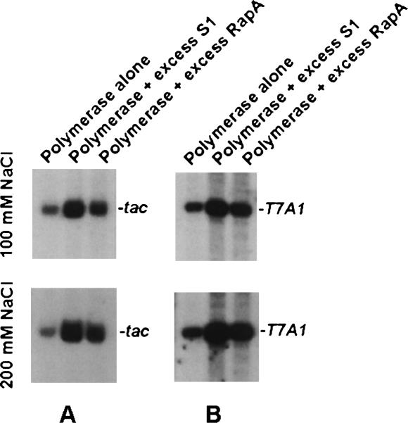
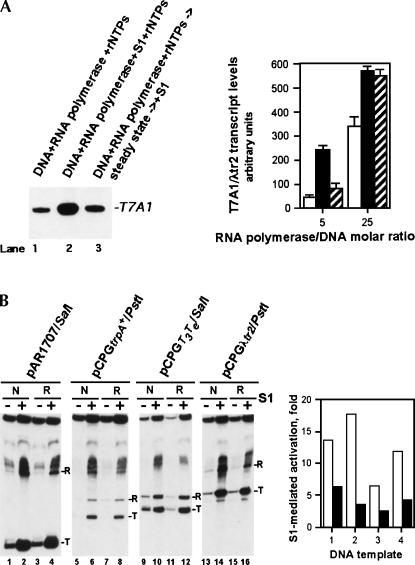
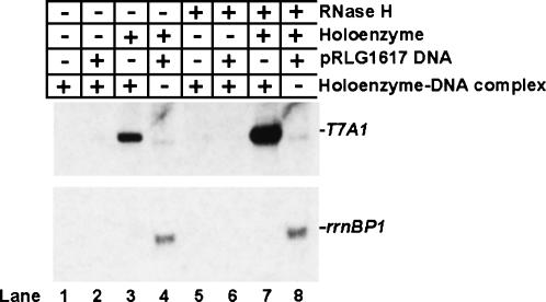
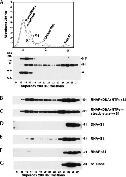
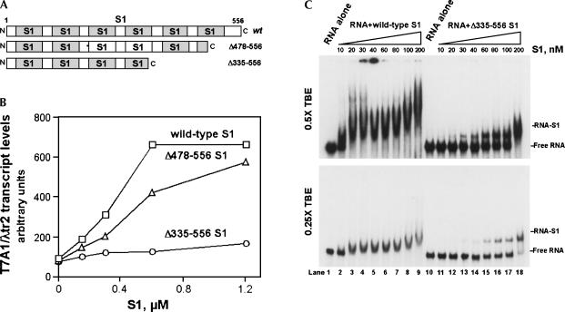
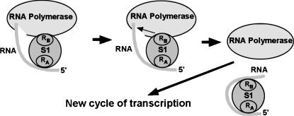
Prokaryotic RNA polymerases are capable of efficient, continuous synthesis of RNA in vivo, yet purified polymerase-DNA model systems for RNA synthesis typically produce only a limited number of catalytic turnovers. Here, we report that the ribosomal protein S1--which plays critical roles in translation initiation and elongation in Escherichia coli and is believed to stabilize mRNA on the ribosome--is a potent activator of transcriptional cycling in vitro. Deletion of the two C-terminal RNA-binding modules--out of a total of six loosely homologous RNA-binding modules present in S1--resulted in a near-loss of the ability of S1 to enhance transcription, whereas disruption of the very last C-terminal RNA-binding module had only a mild effect. We propose that, in vivo, cooperative interaction of multiple RNA-binding modules in S1 may enhance the transcript release from RNA polymerase, alleviating its inhibitory effect and enabling the core enzyme for continuous reinitiation of transcription.


Figures

FIGURE 1.
FIGURE 1.
The ribosomal protein S1 enhances transcriptional cycling. In vitro transcription reactions with supercoiled DNA template pCPGλtr2 (Reynolds et al. 1992) were carried out as described in Materials and Methods. (A) Kinetics of the transcription-stimulatory activity of S1. Reactions were carried out in the presence or absence of 0.2 μM purified native S1. The position of the T7A1 promoter-generated transcript RNA terminated at a specific transcription terminator (λtr2) is indicated. (B) The yields of T7A1/λtr2 transcript plotted as a function of native S1 concentration. In vitro transcription reactions were carried out in the presence of 50mM NaCl (open symbols) and 100 mM NaCl (closed symbols). (C) The transcription-stimulatory effect of S1 is abolished in the presence of high concentrations of the DNA competitor heparin. The reaction conditions were as described in Materials and Methods, with the exception that heparin was added to some of the reactions along with the rNTP premix. In vitro transcription reactions were performed in the absence (open boxes) or in the presence (solid boxes) of 0.2 μM purified native S1.
FIGURE 2.
FIGURE 2.
Back-to-back comparison of the transcription-stimulatory activity of S1 and RapA. Reactions with ptac1617 (Sukhodolets et al. 2001) (A) and pCPGλtr2 (B) supercoiled DNA templates were carried out in the presence of 2.1 μM S1 and 2.4 μM RapA. RNA polymerase/DNA template ratio and concentrations and conditions for in vitro transcription reactions were as described earlier (Sukhodolets et al. 2001). Promoter-specific transcripts resulting from in vitro transcription reactions carried out in the presence of 100 mM NaCl (top) and 200 mM NaCl (bottom) are shown.
FIGURE 3.
FIGURE 3.
Effect of delayed addition of S1 to in vitro transcription reactions and the effect of S1 on the efficiency of transcription termination. (A) The delayed addition of S1 to in vitro transcription reactions results in greatly reduced transcription-stimulatory activity. The quantitated results of two independent, parallel experiments are shown. Reactions in lanes 1 (open columns) and 2 (black columns) are similar to those in lanes 5 and 10 of Figure 1A; in lane 3 (hatched columns), purified native S1 was added 30 min after the initiation of the in vitro transcription reaction, and the reactions were incubated at 37°C for an additional 60 min. The reactions were performed with an approximately fivefold (see Materials and Methods) or 25-fold molar excess of RNA polymerase over the supercoiled pCPGλtr2 DNA template. The RNA polymerase/DNA template molar ratios are indicated. Incubation of RNA polymerase alone at 37°C up to 90 min produced no significant reduction in the enzyme's activity. (B) Mild stimulation of the transcription termination efficiency in the presence of 0.3 μM purified native S1 (lanes “N”) or 0.6 μM recombinant His-tagged S1 (lanes “R”). RNA transcripts terminated at specific terminators (T) and runoff RNA transcripts (R) are indicated. The quantitated results of the experiment are shown on the right; open and black columns show the magnitudes of the S1-mediated transcriptional activation for RNA transcripts terminated at specific terminators and runoff RNA transcripts, respectively. The linearized plasmid DNA templates used in this experiment were pAR1707 (1), pCPGtrpA + (2), pCPGT3Te (3), and pCPGλtr2 (4). DNA templates carried the T7A1 promoter followed by a defined transcription terminator downstream (the type of terminator is apparent from the plasmids' names); all fourtemplates are described in Reynolds et al. (1992).
FIGURE 4.
FIGURE 4.
Inactivation of RNA polymerase rather than the DNA template is the principal reason for poor transcriptional cycling in in vitro transcription reactions with supercoiled DNA. Five microliters of 500 mM Tris-HCl, pH 7.5, 20 mM MgCl2, 1 M NaCl were mixed with 25 μL of purified water, 5 μL RNA polymerase holoenzyme (1.2mg/mL), 5 μL pCPGλtr2 supercoiled DNA (0.89 mg/mL) with the T7A1 promoter and λtr2 terminator, 10 μL of 5× rNTP mix containing 1 mM each of ATP, GTP, CTP, and UTP, and 0.01 μCi of [α32P] UTP. Following a 30-min incubation at 37°C, the entire 50-μL reaction mixture was passed through a Superdex 200 HR 10/30 column pre-equilibrated with TGED buffer containing 100 mM NaCl; 0.5-mL fractions were collected. Aliquots of 250 μL from fractions containing the DNA-bound transcription complexes were concentrated to ∼25 μL using Microcon-10 concentrators (Amicon), and 3-μL aliquots of the resulting purified RNA polymerase–DNA complex were used in in vitro transcription reactions, performed as described in Materials and Methods, with the exception that RNA polymerase and the DNA template were omitted. When indicated, RNA polymerase holoenzyme was added to a final concentration of 0.05 mg/mL; pRLG1617 supercoiled DNA template with the ribosomal rrnBP1 promoter and T1T2 terminator (Ross et al. 1990) to 0.08 mg/mL; and RNase H (New England Biolabs), 5 U per 20 μL reaction. A representative result of two independent experiments is shown.
FIGURE 5.
FIGURE 5.
S1 interacts with transcription complexes. (A) Top: the A280 profile resulting from size-separation of an in vitro transcription reaction mixture on a Superdex 200 HR 10/30 column. The supercoiled pCPGλtr2 DNA template (Reynolds et al. 1992) used in this set of experiments carried the T7A1 promoter and λtr2 terminator. The individual components of the reaction were identified as described in Materials and Methods. Bottom: S1 and RNA polymerase subunits inthe fractions were also identified by Western blot analysis using S1-specific and core RNA polymerase-specific antibodies. (B,C) The in vitro transcription reactions were similar to those described in Materials and Methods, with the exceptions that 0.24× (12 μL) reactions were utilized here, and the reactions contained 0.2 μM purified native S1. The reaction ingredients are indicated at the right. S1 in the Superdex 200 HR 10/30 column fractions is visualized here and below by immunoblotting with S1-specific antibodies. (D) 0.2 μM S1 plus pCPGλtr2 supercoiled DNA (0.089 mg/mL). (E) In vitro transcription reactions similar to those described above were carried out and, after a 30-min incubation at 37°C, 4 U of RNase-free DNase (Promega) was added to the mixtures to digest the DNA template. Following another 30-min incubation at 37°C, the reactions were deproteinized by phenol extraction, and the aqueous phase was precipitated with ethanol. The RNA pellet was redissolved in 12 μL of 1× TB containing 0.2 μM purified native S1, and the binding experiments were performed as described in Materials and Methods. The substitution of DNase with a DNase/RNaseH mixture in the above purification procedure produced similar S1–RNA binding patterns, suggesting that DNA–RNA hybrids are unlikely to play a major role in the interaction of S1 with transcription complexes (data not shown). (F) 0.2 μM S1 plus 1.3 μM RNAP holoenzyme. In a similar binding assay, Δ335–556 S1 showed no detectable complex formation with the polymerase. (G) 0.24 μM S1.
FIGURE 6.
FIGURE 6.
Alteration of the transcription-stimulatory activity of S1 by deletion mutations at the protein's C terminus. (A) Schematic illustrating the modular composition of S1. (B) Effects of C-terminal deletion mutations on the in vitro transcription-stimulatory activity of S1. The transcription-stimulatory activities of the wild-type and mutant S1 proteins are plotted as a function of protein concentration. In vitro transcription reactions were carried out as described in Materials and Methods. (C) The RNA-binding activity of wild-type S1 versus that of Δ335–556 S1. The gels show titrations of purified labeled T7A1/λtr2 RNA with purified recombinant S1 proteins; the S1 concentrations are indicated. EMSA experiments were carried out as described in Materials and Methods.
FIGURE 7.
FIGURE 7.
Cooperative binding of RNA polymerase-associated transcript RNA by multiple RNA-binding modules in S1 may effectively strip transcript from RNA polymerase, alleviating its inhibitory effect. RA and RB denote independent RNA-binding segments in S1 which may incorporate one or more individual RNA-binding or “S1”-modules (defined as described in Bycroft etal. 1997).

References

    1. Arndt K.M., Chamberlin M.J. Transcription termination in Escherichia coli. Measurement of the rate of enzyme release from Rho-independent terminators. J. Mol. Biol. 1988;202:271–285. - PubMed
    1. Berlin V., Yanofsky C. Release of transcript and template during transcription termination at the trp operon attenuator. J.Biol. Chem. 1983;258:1714–1719. - PubMed
    1. Boni I.V., Isaeva D.M., Musychenko M.L., Tzareva N.V. Ribosome-messenger recognition: mRNA target sites for ribosomal protein S1. Nucleic Acids Res. 1991;19:155–162. - PMC - PubMed
    1. Boni I.V., Artamonova V.S., Tzareva N.V., Dreyfus M. Non-canonical mechanism for translational control in bacteria: Synthesis of ribosomal protein S1. EMBO J. 2001;20:4222–4232. - PMC - PubMed
    1. Bycroft M., Hubbard T.J.P., Proctor M., Freund S.M.V., Murzin A.G. The solution structure of the S1 RNA binding domain: A member of an ancient nucleic acid-binding fold. Cell. 1997;88:235–242. - PubMed

Publication types

MeSH terms

LinkOut - more resources