. 2006 Jul;80(13):6276-85.
doi: 10.1128/JVI.00147-06.

Characterization of the RNA chaperone activity of hantavirus nucleocapsid protein

Affiliations

Characterization of the RNA chaperone activity of hantavirus nucleocapsid protein

M A Mir et al. J Virol. 2006 Jul.

Abstract

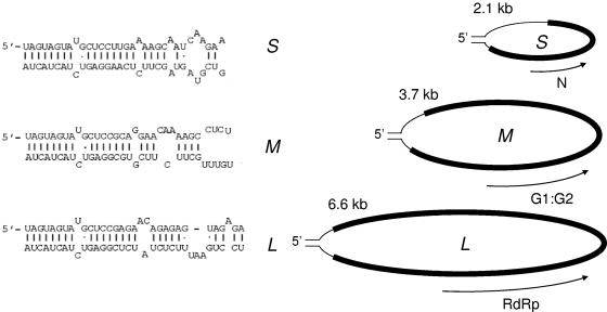
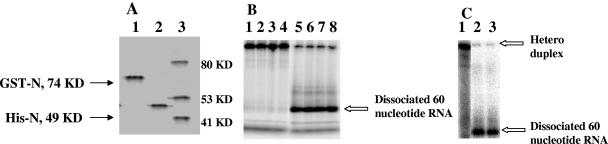
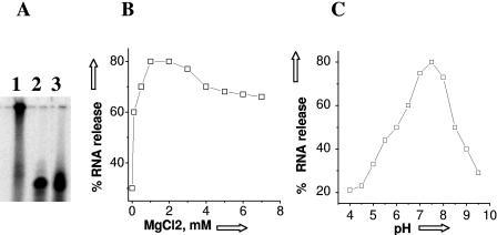
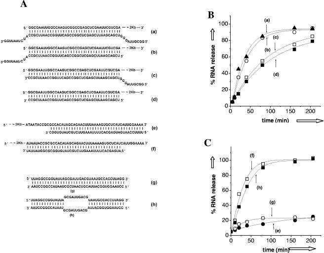
Hantaviruses are tripartite negative-sense RNA viruses and members of the Bunyaviridae family. The nucleocapsid (N) protein, encoded by the smallest of the three genome segments (S), has nonspecific RNA chaperone activity. This activity results in transient dissociation of misfolded RNA structures, may be required for facilitating correct higher-order RNA structure, and may function in viral genome replication. We carried out a series of experiments to further characterize the ability of N to dissociate RNA duplexes. As might be expected, N dissociated RNA duplexes but not DNA duplexes or RNA-DNA heteroduplexes. The RNA-destabilizing activity of N is ATP independent, has a pH optimum of 7.5, and has an Mg(2+) concentration optimum of 1 to 2 mM. N protein is unable to unwind the RNA duplexes that are completely double stranded. However, in the presence of an adjoining single-stranded region, helix unwinding takes place in the 3'-to-5' direction through an unknown mechanism. The N protein trimer specifically recognizes and unwinds the terminal panhandle structure in the viral RNA and remains associated with unwound 5' terminus. We suggest that hantaviral nucleocapsid protein has an active role in hantaviral replication by working cooperatively with viral RNA polymerase. After specific recognition of the panhandle structure by N protein, the unwound 5' terminus likely remains transiently bound to N protein, creating an opportunity for the viral polymerase to initiate transcription at the accessible 3' terminus.


Figures

FIG. 1.
FIG. 1.
Tripartite hantaviral genome. The sequences of the 5′ and 3′ termini that form the general “panhandle” structure of the S, M, and L segments are shown.
FIG. 2.
FIG. 2.
RNA helix dissociation activity of SNV N protein. (A) Purified SNV N protein expressed and purified from E. coli containing an N-terminal GST tag (lane 1) or a C-terminal His6 tag (lane 2). Protein molecular mass markers are shown in lane 3. The theoretical molecular masses of GST-N protein and His6-tagged N proteins are indicated, and the mobility of these N derivatives corresponds well to the size standards. (B) RNA helix dissociation assays. Heteroduplexes were formed as described in Materials and Methods. Lane 1 shows a gel-purified heteroduplex. Lane 2, an aliquot from a 20-μl helix dissociation reaction containing a 1:1 RNA/protein ratio (75 nM heteroduplex RNA and 75 nM BSA); lane 3, 75 nM heteroduplex RNA and 75 nM human tissue factor pathway inhibitor-2 (TFPI-2); lane 4, 75 nM heteroduplex RNA and 75 nM biotin protein ligase; lane 5, heteroduplex heated at 95°C for 5 min before loading into gel; lane 6, 75 nM RNA heteroduplex and 75 nM GST-N; lane 7, 75 nM heteroduplex RNA and 75 nM His6-N protein; and lane 8, 75 nM heteroduplex RNA and 75 nM HIV-1 His-Gag. RNAs were fractionated by 12% SDS-polyacrylamide gel electrophoresis (SDS-PAGE). (C) Comparison of the helix dissociation assays of purified trimeric N and unfractionated N. Lane 1, a 12% SDS gel with purified heteroduplex; lane 2, heteroduplex preincubated with unfractionated N; lane 3, heteroduplex preincubated with purified trimeric N.
FIG. 3.
FIG. 3.
Effect of ATP, MgCl2, and pH on the RNA helix dissociation activity of SNV N. (A) Effect of ATP on N-mediated helix dissociation. Lane 1 contains heteroduplex RNA incubated without added N protein, lane 2 contains the products of a reaction that contained RNA heteroduplex (as in Fig. 2) and N protein in the presence of 1 mM ATP, and lane 3 contains the products of a reaction without ATP. (B) Effect of MgCl2 on helix dissociation. The graph shows the percent release of the radiolabeled shorter RNA from heteroduplex RNA by N at a range of MgCl2 concentrations. (C) Effect of pH on the helix dissociation activity of N. The graph shows the percent release of the radiolabeled shorter RNA from the RNA heteroduplex as a function of pH.
FIG. 4.
FIG. 4.
Helix dissociation activity of SNV N is specific for heteroduplexes composed solely of RNA. (A) Kinetics of N-mediated helix dissociation using a heteroduplex with both the longer and shorter nucleic acid composed of RNA [heteroduplex (a) of panel B]. The RNA heteroduplex was incubated with N protein for increasing lengths of time and fractionated on a 12% gel. t = 0, lane 1; t = 10 min, lane 2; t = 20 min, lane 3; t = 40 min, lane 4; t = 80 min, lane 5; t = 160 min, lane 6; and t = 205 min, lane 7. (B) Various RNA and DNA heteroduplexes used in helix dissociation assays. (a) Heteroduplex formed between a longer RNA molecule and a small, radiolabeled RNA 60 nucleotides long. (b) Heteroduplex containing the same “longer” RNA as in panel a but with a shorter labeled DNA. (c) Heteroduplex containing a longer DNA and the same shorter RNA as in panel a. (d) Heteroduplex composed solely of DNA. (C) N-mediated dissociation of the various heteroduplexes shown in panel B. Samples from helix dissociation reactions were fractionated on 12% SDS-PAGE and exposed to phosphorimager screen, as in panel A, and dissociation was quantified using phosphorimaging.
FIG. 5.
FIG. 5.
N-mediated helix dissociation requires a single-stranded region 3′ to the duplex. (A) Several related RNA heteroduplexes were generated to characterize N-mediated helix dissociation. These include a molecule containing a shorter RNA complementary to the 5′ terminus of a longer RNA with an additional 10 noncomplementary nucleotides at both the 5′ and 3′ ends (a). (b) A molecule similar to that in panel a but the 5′ end of the shorter RNA is exactly complementary to the longer RNA. (c) The molecule is identical to that in panel a except that the 3′ end of the shorter RNA is exactly complementary to the longer RNA. (d) A molecule similar to that in panel a except that both the 5′ and 3′ ends of the shorter RNA are exactly complementary to the longer RNA. As shown in the diagram, the shorter RNA of the molecules shown in panels a through d is complementary to the 5′ end of the longer RNA. In contrast, in panels e and f, the shorter RNA is complementary to the region at or near the 3′ end of the longer RNA. The heteroduplex in panel e is a 40-nucleotide radiolabeled short RNA complementary to the 3′ end of the longer RNA, whereas that in panel f contains a smaller RNA that is complementary to the longer RNA beginning at a site 10 nucleotides from the 3′ end of the longer RNA. (g) Heteroduplex composed of two relatively small RNAs (40 nucleotides in length) that are exactly complementary to each other. (h) Heteroduplex similar to that in panel g except that the central 10 bases of the heteroduplex are not complementary. (B) Kinetic dissociation profiles for heteroduplexes shown in panels a, b, c, and d of panel A. (C) Kinetic dissociation profiles for heteroduplexes shown in panels e, f, g, and h from panel A.
FIG. 6.
FIG. 6.
N-mediated dissociation of helices corresponding to the viral panhandle. (A) The heteroduplex formed by 32 nucleotides from 3′ and 5′ ends of the SNV S segment RNA. In panel i, the 3′ nucleotides were radioactively labeled, whereas in panel ii the 5′ nucleotides were labeled. Labeled nucleotides are indicted by bold lettering. (B) Kinetics for the dissociation of radiolabeled 3′ and 5′ nucleotides from the heteroduplexes shown in panels i and ii. (C) RNA filter binding data using the two heteroduplexes. Each heteroduplex was incubated with N protein trimer for the indicated time period, and protein-dependent retention of radiolabeled 3′ nucleotides of the panhandle (shown by dark boxes) and 5′ nucleotides of the panhandle (shown by open boxes) was measured. The molar ratio of RNA/N was 1:1. Panel D shows the results of an RNA filter binding assay with increasing concentrations of N protein trimer and either the 3′-terminal 32 nucleotides (open squares) or the 5′-terminal 32 nucleotides (filled squares). Measured dissociation constants for both RNAs are also shown. N protein was incubated with radiolabeled RNA for 45 min at room temperature before filtration through nitrocellulose filters.
FIG. 7.
FIG. 7.
Reverse transcription using SNV S minipanhandle as a template. Panel A depicts an SNV S segment minipanhandle containing 32 nucleotides from both the 3′ and 5′ end of S segment RNA separated by six uracil residues that comprise the loop sequence. An RNA primer complementary to the 3′ end of this minipanhandle, which was used as a primer for reverse transcription, is also shown at the bottom of the minipanhandle. (B) Reverse transcription reactions using SNV minipanhandle and the RNA primer shown in panel A. SNV S minipanhandle template and RNA primer incubated at RT without thermal denaturation or addition of N protein are shown in lane 1. The SNV S minipanhandle template and RNA primer incubated at RT with N protein and without thermal denaturation are shown in lane 2. Reverse transcription products from a reaction with the SNV S minipanhandle template and RNA primer following thermal denaturation and primer annealing are shown in lane 3. (C) Reverse transcription products produced at different molar ratios of trimeric N and the SNV S minipanhandle in the absence of thermal denaturation. Samples were fractionated using 12% SDS-PAGE in panel B, whereas an 8% gel was used in panel C.

Similar articles

Cited by

References

    1. Alfadhli, A., Z. Love, B. Arvidson, J. Seeds, J. Willey, and E. Barklis. 2001. Hantavirus nucleocapsid protein oligomerization. J. Virol. 75:2019-2023. - PMC - PubMed
    1. Alfadhli, A., E. Steel, L. Finlay, H. P. Bachinger, and E. Barklis. 2002. Hantavirus nucleocapsid protein coiled-coil domains. J. Biol. Chem. 277:27103-27108. - PubMed
    1. Blakqori, G., G. Kochs, O. Haller, and F. Weber. 2003. Functional L polymerase of La Crosse virus allows in vivo reconstitution of recombinant nucleocapsids. J. Gen. Virol. 84:1207-1214. - PubMed
    1. Bridgen, A., and R. M. Elliott. 1996. Rescue of a segmented negative-strand RNA virus entirely from cloned complementary DNAs. Proc. Natl. Acad. Sci. USA 93:15400-15404. - PMC - PubMed
    1. Cristofari, G., R. Ivanyi-Nagy, C. Gabus, S. Boulant, J. P. Lavergne, F. Penin, and J. L. Darlix. 2004. The hepatitis C virus Core protein is a potent nucleic acid chaperone that directs dimerization of the viral (+) strand RNA in vitro. Nucleic Acids Res. 32:2623-2631. - PMC - PubMed

Publication types

MeSH terms