. 2006 Jul;80(13):6286-94.
doi: 10.1128/JVI.02648-05.

Herpes simplex virus capsid structure: DNA packaging protein UL25 is located on the external surface of the capsid near the vertices

Affiliations

Herpes simplex virus capsid structure: DNA packaging protein UL25 is located on the external surface of the capsid near the vertices

William W Newcomb et al. J Virol. 2006 Jul.

Abstract

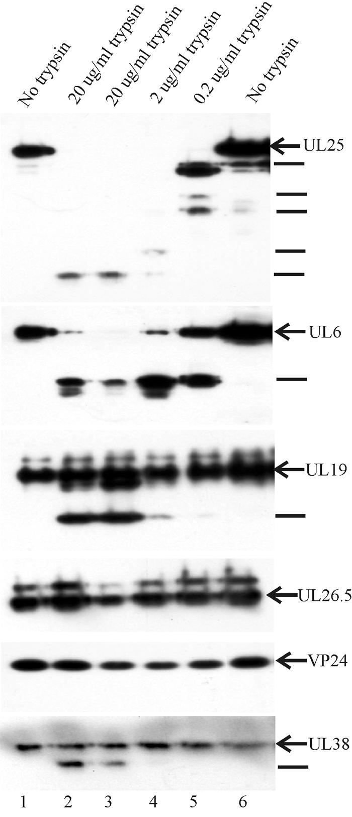
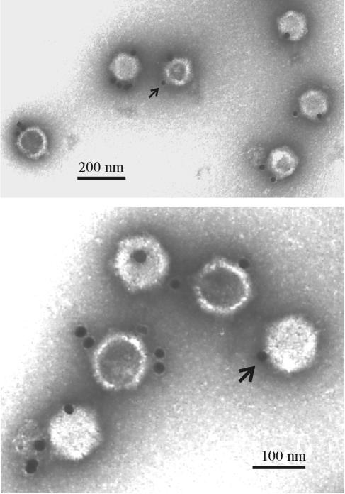
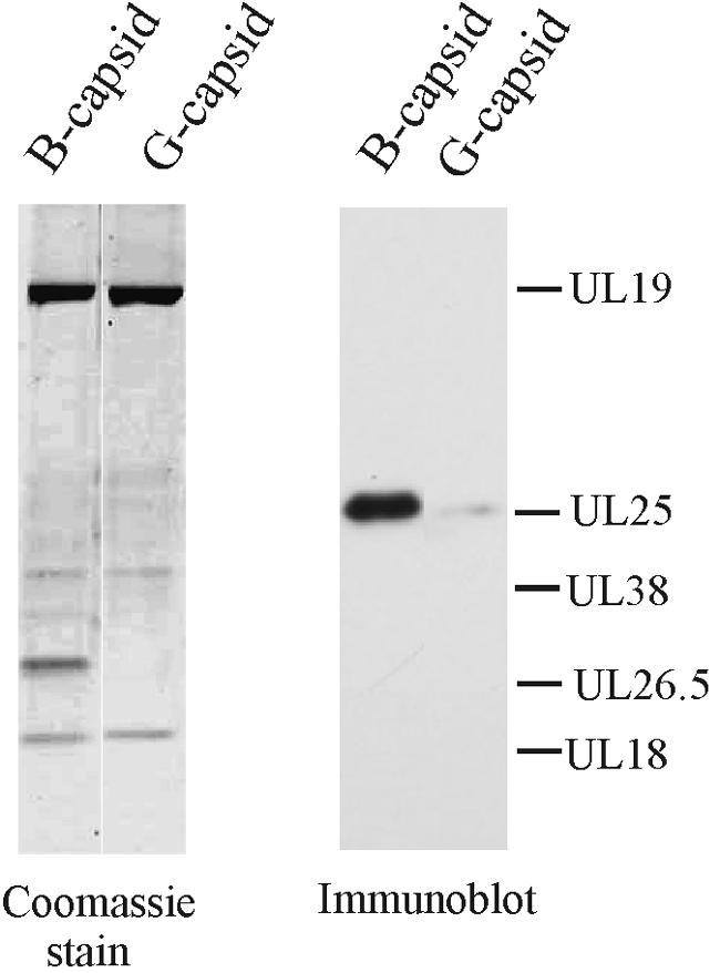
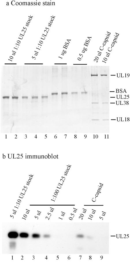
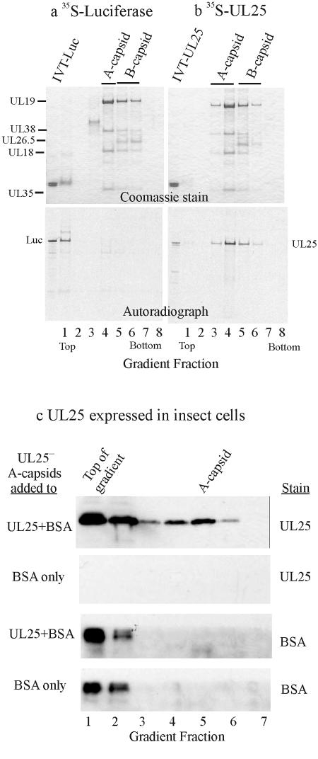
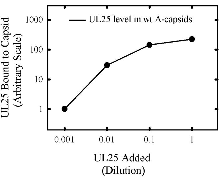
UL25 is one of seven herpes simplex virus-encoded proteins involved specifically in DNA encapsidation. Its role appears to be to stabilize the capsid so that DNA is prevented from escaping once it has entered. To clarify the function of UL25, we have examined capsids with the goal of defining where it is located. Analysis of trypsin-treated capsids showed that UL25 is sensitive to cleavage like other proteins such as the major capsid and portal proteins that are exposed on the capsid surface. Internal proteins such as the scaffolding protein and protease were not affected under the same experimental conditions. Capsids were also examined by electron microscopy after staining with gold-labeled antibody specific for UL25. Images of stained capsids demonstrated that most labeled sites (71% in C capsids) were at capsid vertices, and most stained C capsids had label at more than one vertex. A quantitative immunoblotting method showed that the capsid contents of UL25 were 56, 20, and 75 copies per capsid in A, B, and C capsids, respectively. Finally, soluble UL25 protein was found to bind in vitro to purified capsids lacking it. The amount of bound UL25 corresponded to the amount present in B capsids, and bound UL25 was found by immunoelectron microscopy to be located predominantly at the capsid vertices. The results are interpreted to suggest that five UL25 molecules are found at or near each of the capsid vertices, where they are exposed on the capsid surface. Exposure on the surface is consistent with the view that UL25 is added to the capsid as DNA is packaged or during late stages of the packaging process.


Figures

FIG. 1.
FIG. 1.
Immunoblot showing the extent to which UL25 and other HSV-1 capsid proteins are cleaved following treatment of B capsids with trypsin. Capsids were treated in vitro with the trypsin doses indicated at the top, purified, and subjected to SDS-PAGE followed by immunoblotting. The same blot was probed sequentially for all six proteins indicated at the right. Lines at the right indicate the positions of individual digestion products. Note evidence of digestion in the case of UL25, UL6, UL19, and UL38, but not UL26.5 or VP24.
FIG. 2.
FIG. 2.
Electron micrographs showing HSV-1 C capsids after staining with antibody specific for UL25 followed by an antiantibody conjugated to gold beads. Note that most gold beads are found at capsid vertices (one is indicated by an arrow in each micrograph) and several capsids have label at more than one site.
FIG. 3.
FIG. 3.
Immunoblot analysis of UL25 in B capsids and in B capsids after treatment with 2 M guanidine-HCl (G capsids). Sister SDS-PAGE gels were stained with Coomassie blue (left) and with antibody specific for UL25 (right). Note that nearly all UL25 is removed by exposure to guanidine-HCl, a procedure that causes removal of capsid pentons (23).
FIG. 4.
FIG. 4.
SDS-polyacrylamide gel electrophoresis (a) and immunoblot analyses (b) used to estimate the UL25 copy number in HSV-1 C capsids. Similar experiments were done with A and B capsids. Known amounts of BSA (a, lanes 6 to 9) were used as standards to estimate the protein concentrations of a UL25 stock solution (a, lanes 1 to 5) and HSV-1 C capsids (a, lanes 10 and 11). The immunoblot signals from known UL25 concentrations (b, lanes 1 to 6) were then used to measure the amount of UL25 present in C capsids (b, lanes 7 to 9).
FIG. 5.
FIG. 5.
Analysis of UL25-negative capsids after exposure to UL25 in vitro. Pooled KUL25NS A and B capsids were allowed to bind [35S]Met-labeled UL25 and then purified by sucrose gradient centrifugation. Gradient fractions were then analyzed by SDS-PAGE followed by Coomassie staining (a and b, top panels) and autoradiography (bottom panels). Note that both A and B capsids bound UL25 (b) whereas neither bound the control protein luciferase (Luc) (a). IVT, in vitro transcription-translation. (c) Results obtained when KUL25NS A capsids were allowed to bind UL25 present in insect cell extracts. After incubation to permit binding to occur, capsids were purified by sucrose gradient centrifugation. Gradient fractions were then analyzed by SDS-PAGE followed by immunoblotting for UL25 (top panels) or BSA (bottom panels). All three analytical procedures were performed with the same blot. Note that capsids bound UL25 but not the control protein BSA.
FIG. 6.
FIG. 6.
Effect of UL25 concentration on the amount bound to UL25-negative capsids. UL25 synthesized in insect cells was added to KUL25NS capsids and incubated as described in Materials and Methods. Capsids were then isolated by sucrose gradient centrifugation, and the amount of UL25 present was determined by immunoblotting. The amount of UL25 present in wild-type (wt) 17MP A capsids was estimated from capsids isolated in a companion sucrose gradient and analyzed on the same blot.

References

    1. Aebi, A., R. van Driel, R. K. L. Bijlenga, B. ten Heggeler, R. van der Broek, A. C. Steven, and P. R. Smith. 1977. Capsid fine structure of T-even bacteriophages. Binding and localization of two dispensable capsid proteins into the P23 surface lattice. J. Mol. Biol. 110:687-698. - PubMed
    1. Al-Kobaisi, M. F., F. J. Rixon, I. McDougall, and V. G. Preston. 1991. The herpes simplex virus UL33 gene product is required for the assembly of full capsids. Virology 180:380-388. - PubMed
    1. Bowman, B. R., R. L. Welschhaus, H. Jayaram, N. D. Stow, V. G. Preston, and F. A. Quiocho. 2006. Structural characterization of the UL25 DNA-packaging protein from herpes simplex virus type 1. J. Virol. 80:2309-2317. - PMC - PubMed
    1. Brown, J. C., M. A. McVoy, and F. L. Homa. 2002. Packaging DNA into herpesvirus capsids, p. 111-153. In A. Holzenburg and E. Bogner (ed.), Structure-function relationships of human pathogenic viruses. Kluwer Academic/Plenum Publishers, London, United Kingdom.
    1. Chang, Y. E., A. P. Poon, and B. Roizman. 1996. Properties of the protein encoded by the UL32 open reading frame of herpes simplex virus type 1. J. Virol. 70:3938-3946. - PMC - PubMed

Publication types

MeSH terms

LinkOut - more resources