. 2006 Jul;80(13):6295-304.
doi: 10.1128/JVI.02381-05.

Influenza virus evades innate and adaptive immunity via the NS1 protein

Affiliations

Influenza virus evades innate and adaptive immunity via the NS1 protein

Ana Fernandez-Sesma et al. J Virol. 2006 Jul.

Abstract

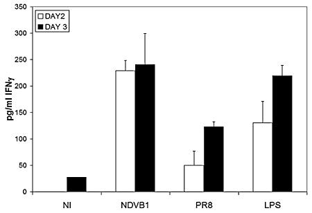
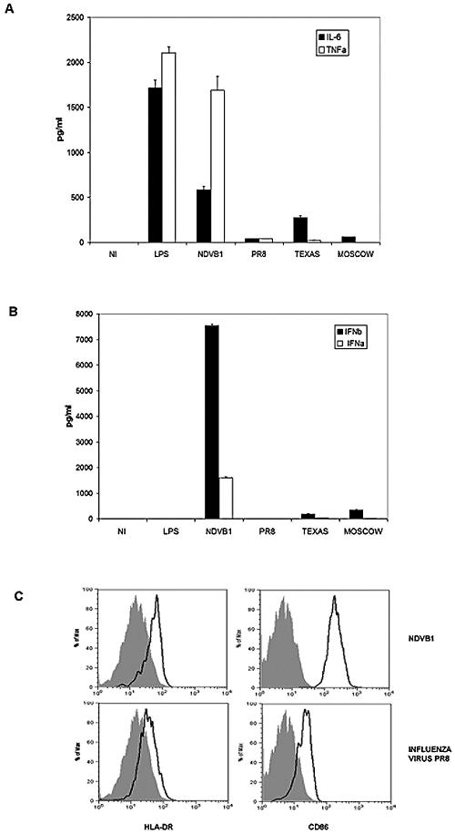
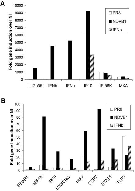
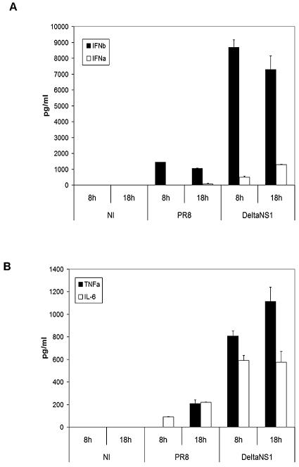
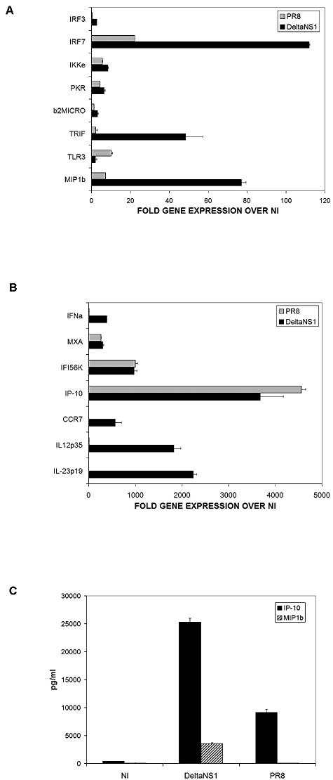
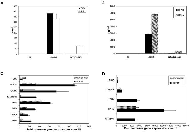
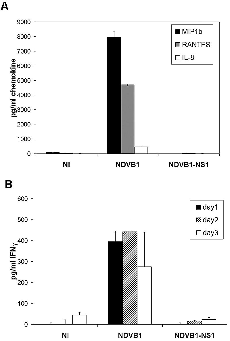
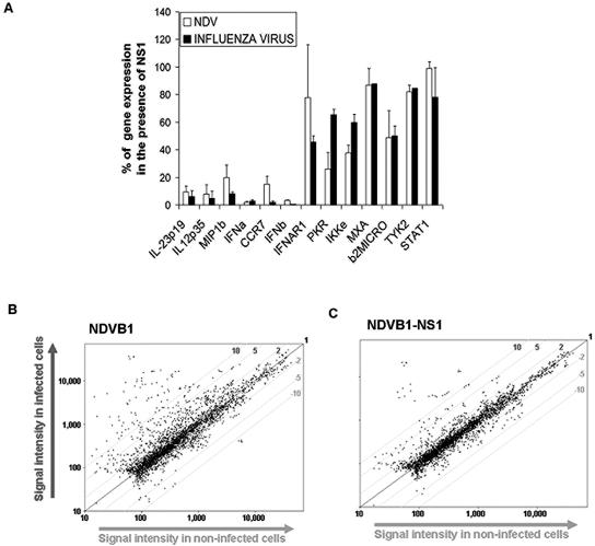
Both antibodies and T cells contribute to immunity against influenza virus infection. However, the generation of strong Th1 immunity is crucial for viral clearance. Interestingly, we found that human dendritic cells (DCs) infected with influenza A virus have lower allospecific Th1-cell stimulatory abilities than DCs activated by other stimuli, such as lipopolysaccharide and Newcastle disease virus infection. This weak stimulatory activity correlates with a suboptimal maturation of the DCs following infection with influenza A virus. We next investigated whether the influenza A virus NS1 protein could be responsible for the low levels of DC maturation after influenza virus infection. The NS1 protein is an important virulence factor associated with the suppression of innate immunity via the inhibition of type I interferon (IFN) production in infected cells. Using recombinant influenza and Newcastle disease viruses, with or without the NS1 gene from influenza virus, we found that the induction of a genetic program underlying DC maturation, migration, and T-cell stimulatory activity is specifically suppressed by the expression of the NS1 protein. Among the genes affected by NS1 are those coding for macrophage inflammatory protein 1beta, interleukin-12 p35 (IL-12 p35), IL-23 p19, RANTES, IL-8, IFN-alpha/beta, and CCR7. These results indicate that the influenza A virus NS1 protein is a bifunctional viral immunosuppressor which inhibits innate immunity by preventing type I IFN release and inhibits adaptive immunity by attenuating human DC maturation and the capacity of DCs to induce T-cell responses. Our observations also support the potential use of NS1 mutant influenza viruses as live attenuated influenza virus vaccines.


Figures

FIG. 1.
FIG. 1.
Allospecific responses of purified CD4 T cells primed with differentially treated DCs. Human DCs were infected with PR8 influenza virus or Newcastle disease virus (NDVB1), left untreated (NI), or treated with 100 ng/ml of LPS for 45 min. Differentially treated DCs were then incubated with allogeneic naïve CD4 T cells for 3 days. Supernatants from the cocultures were harvested at different days of culture and tested by ELISA for the release of IFN-γ into the supernatants of the cocultures. Error bars are for triplicate samples. These results are representative of three independent experiments.
FIG. 2.
FIG. 2.
Influenza virus and NDV induce different degrees of maturation in human DCs. Human DCs were infected on day 5 or 6 of culture with influenza virus (PR8, Texas, or Moscow) or NDV (NDVB1) at an MOI of 0.5. Supernatants from infected cells (at 18 h postinfection) were tested by ELISA for the release of (A) TNF-α and IL-6 and (B) IFN-α and IFN-β. (C) After infection, cells were incubated at 37°C for 18 h and stained for flow cytometry analysis of the expression of HLA-DR (left panels) or CD86 (right panels). Filled histograms represent uninfected cells, and open histograms represent infected cells (with NDVB1 [top panels] and influenza virus PR8 [bottom panels]). Concentrations are indicated in pg/ml. Error bars represent the standard errors of triplicate samples, and data are representative of at least three independent experiments with cells from different donors.
FIG. 3.
FIG. 3.
Infection of human DCs with influenza virus PR8 resulted in low expression of genes involved in DC maturation compared with that in NDV infection. Human DCs were either infected with NDVB1 or influenza virus PR8 at an MOI of 0.5 or treated with 5,000 U/ml of IFN-β or 500 ng/ml of LPS. At different times after treatment, RNAs were isolated and used to generate cDNAs to test increases in specific gene expression. (A) Genes upregulated at high levels 24 h after treatment; (B) genes upregulated at moderate levels 24 h after treatment. The data are representative of at least three independent experiments with cells from different donors and are given as x-fold increases in gene expression over that in uninfected cells (NI).
FIG. 4.
FIG. 4.
The NS1 protein of influenza virus abolishes the release of proinflammatory cytokines and IFN-α/β by human DCs after infection with influenza virus. Human DCs were infected with influenza virus PR8 or DeltaNS1 or mock infected. Supernatants from infected DCs were tested by ELISA for the release of (A) IFN-α (white bars) and IFN-β (black bars) or (B) TNF-α (black bars) and IL-6 (white bars) at different times after infection. Error bars are for triplicate samples. Data are representative of at least three independent experiments.
FIG. 5.
FIG. 5.
The NS1 protein of influenza virus downregulates the expression of specific genes involved in DC maturation. Human DCs were infected with influenza viruses PR8 and DeltaNS1 for 18 h. (A and B) RNAs were isolated from the cells and used to generate cDNAs to perform qRT-PCR for genes involved in DC maturation. Values indicate changes in gene expression in DCs infected by viruses compared to that in uninfected DCs (NI). Error bars represent standard deviations for triplicate samples. Results are representative of three independent experiments with cells from three different donors. (C) At 18 h postinfection, supernatants from infected DCs were analyzed by ELISA for the presence of IP-10 and MIP1β. Error bars are for triplicate samples. Results are representative of three independent experiments with cells from three different donors.
FIG. 6.
FIG. 6.
The NS1 protein of influenza virus downregulates DC maturation when expressed in an NDV background. Human DCs were infected with NDVB1 and NDVB1-NS1 for 18 h. Supernatants from infected DCs were analyzed by ELISA for the presence of (A) TNF-α and IL-6 or (B) IFN-α and IFN-β. Error bars are for triplicate samples. Results are representative of three independent experiments with cells from three different donors. (C and D) RNAs were isolated from the cells and used to perform qRT-PCR for genes involved in DC maturation. Values indicate changes in gene expression in DCs infected by viruses compared to that in uninfected DCs (NI). Error bars represent standard deviations for triplicate samples. Results are representative of three independent experiments with cells from three different donors.
FIG. 7.
FIG. 7.
The NS1 protein of influenza virus has an inhibitory effect on chemokine secretion and T-cell priming by DCs. (A) Supernatants from DCs infected with NDVB1 or NDVB1-NS1 or not infected (NI) were tested by ELISA for the presence of MIP1β, RANTES, and IL-8. Error bars are for triplicate samples. Data are representative of at least three independent experiments. (B) Human DCs were infected with NDVB1 or NDVB1-NS1 at an MOI of 0.5 for 45 min and then incubated with peripheral blood mononuclear cells from an allogeneic donor at a ratio of 1:1 for 3 days. Supernatants were harvested every day and tested by ELISA for the release of IFN-γ. Results represent three independent experiments. Error bars are for triplicate samples in each experiment.
FIG. 8.
FIG. 8.
The inhibitory effect of the NS1 protein on DC maturation is not a global effect. (A) Human DCs were infected with influenza virus PR8 or DeltaNS1 or with NDVB1 or NDVB1-NS1 for 18 h. RNAs were isolated from the cells and used to perform qRT-PCR for genes involved in DC maturation. White bars represent effects of NDVB1-NS1 over those of NDVB1 (NDV), while black bars represent effects of PR8 influenza virus over those of DeltaNS1 (influenza virus). Values indicate percentages of gene expression by viruses expressing NS1 relative to that by the same viruses lacking NS1. Error bars represent standard deviations from three independent experiments with cells from three different donors. (B and C) Human DCs infected with NDVB1 and NDVB1-NS1 were analyzed by microarray analysis against uninfected DCs, using an immunology microarray panel of 1,070 genes (Memorec; Miltenyi Biotec). y axes show signal intensities in infected cells, and x axes show signal intensities in uninfected cells.

References

    1. Alcami, A., and U. H. Koszinowski. 2000. Viral mechanisms of immune evasion. Trends Microbiol. 8:410-418. - PMC - PubMed
    1. Banchereau, J., F. Briere, C. Caux, J. Davoust, S. Lebecque, Y. J. Liu, B. Pulendran, and K. Palucka. 2000. Immunobiology of dendritic cells. Annu. Rev. Immunol. 18:767-811. - PubMed
    1. Banchereau, J., and R. M. Steinman. 1998. Dendritic cells and the control of immunity. Nature 392:245-252. - PubMed
    1. Barchet, W., A. Krug, M. Cella, C. Newby, J. A. Fischer, A. Dzionek, A. Pekosz, and M. Colonna. 2005. Dendritic cells respond to influenza virus through TLR7- and PKR-independent pathways. Eur. J. Immunol. 35:236-242. - PubMed
    1. Belladonna, M. L., J. C. Renauld, R. Bianchi, C. Vacca, F. Fallarino, C. Orabona, M. C. Fioretti, U. Grohmann, and P. Puccetti. 2002. IL-23 and IL-12 have overlapping, but distinct, effects on murine dendritic cells. J. Immunol. 168:5448-5454. - PubMed

Publication types

MeSH terms