Comparative Study
. 2006 Jul;80(13):6368-77.
doi: 10.1128/JVI.00211-06.

Visualization of intracellular transport of vesicular stomatitis virus nucleocapsids in living cells

Affiliations
Comparative Study

Visualization of intracellular transport of vesicular stomatitis virus nucleocapsids in living cells

Subash C Das et al. J Virol. 2006 Jul.

Abstract

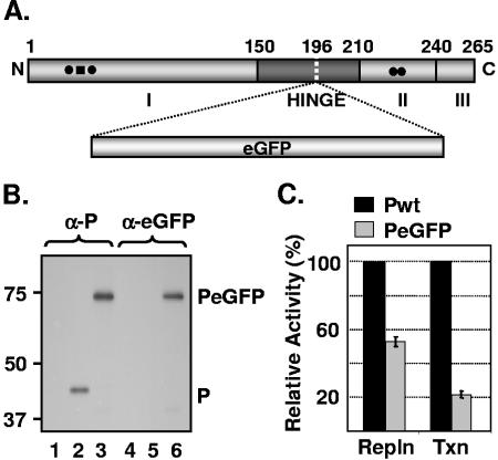
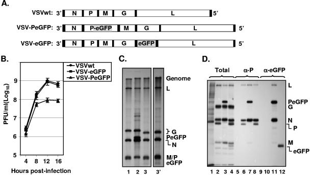
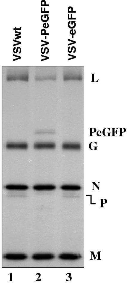
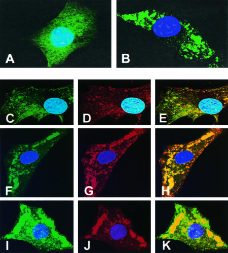
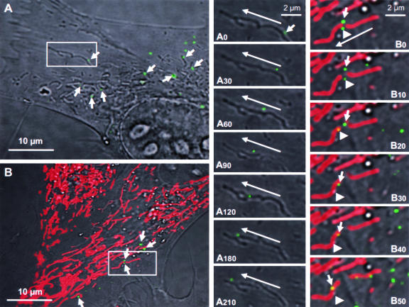
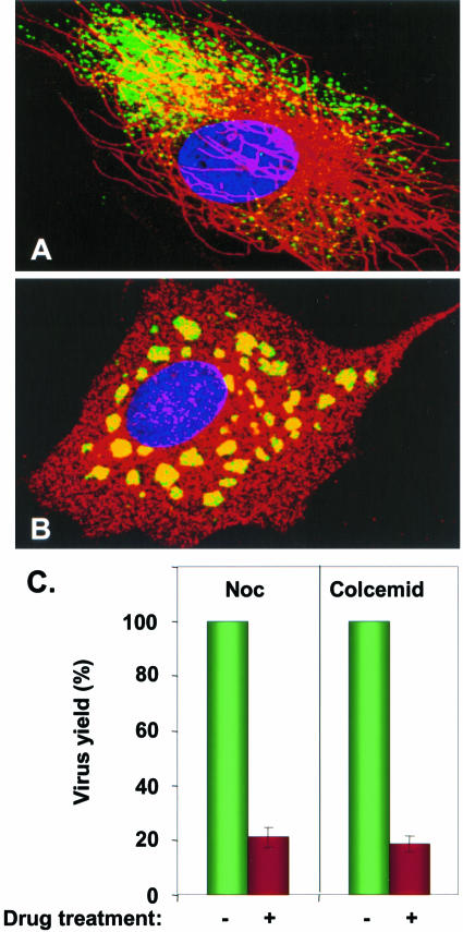
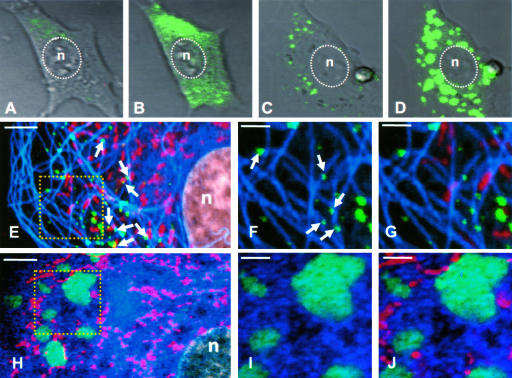
The phosphoprotein (P) of vesicular stomatitis virus (VSV) is a subunit of the viral RNA polymerase. In previous studies, we demonstrated that insertion of 19 amino acids in the hinge region of the protein had no significant effect on P protein function. In the present study, we inserted full-length enhanced green fluorescent protein (eGFP) in frame into the hinge region of P and show that the fusion protein (PeGFP) is functional in viral genome transcription and replication, albeit with reduced activity. A recombinant vesicular stomatitis virus encoding PeGFP in place of the P protein (VSV-PeGFP), which possessed reduced growth kinetics compared to the wild-type VSV, was recovered. Using the recombinant VSV-PeGFP, we show that the viral replication proteins and the de novo-synthesized RNA colocalize to sites throughout the cytoplasm, indicating that replication and transcription are not confined to any particular region of the cytoplasm. Real-time imaging of the cells infected with the eGFP-tagged virus revealed that, following synthesis, the nucleocapsids are transported toward the cell periphery via a microtubule (MT)-mediated process, and the nucleocapsids were seen to be closely associated with mitochondria. Treatment of cells with nocodazole or Colcemid, drugs known to inhibit MT polymerization, resulted in accumulation of the nucleocapsids around the nucleus and also led to inhibition of infectious-virus production. These findings are compatible with a model in which the progeny viral nucleocapsids are transported toward the cell periphery by MT and the transport may be facilitated by mitochondria.


Figures

FIG. 1.
FIG. 1.
Replication and transcription activities of PeGFP fusion protein. (A) Domain organization of P protein, showing domains I, II, and III and the hinge region. The site of eGFP incorporation at aa position 196 is indicated by a vertical dotted line. (B) Expression of PeGFP fusion protein in transfected cells. Cells transfected with plasmids encoding P (lanes 2 and 5) or PeGFP (lanes 3 and 6) proteins or no plasmid (lanes 1 and 4) were radiolabeled with Expre35S35S label. The radiolabeled proteins were immunoprecipitated with antibodies as shown on the top (α-P, anti-P; α-eGFP, anti-eGFP), analyzed by SDS-PAGE, and detected by fluorography. Size markers in kDa are shown on the left. P and PeGFP proteins are identified on the right. (C) Replication and transcription activities of PeGFP protein relative to Pwt as determined by DI-particle replication or minigenome transcription assays (20, 40). The histograms represent the average data from three independent experiments, with standard deviations shown by error bars. Repln, replication; Txn, transcription.
FIG. 2.
FIG. 2.
Recovery and characterization of recombinant VSV encoding PeGFP fusion protein. (A) Recombinant VSV genome plasmids. VSVwt, the wt VSV genome with the N, P, M, G, and L genes, shown in rectangular boxes; VSV-PeGFP, the VSV genome containing the PeGFP gene in place of the wt P gene; VSV-eGFP, the VSV genome containing the eGFP gene as an extra cistron. Intergenic regions as well as 3′ leader gene and 5′ trailer sequences are shown in black boxes; the eGFP coding region is shaded. (B) Single-cycle growth kinetics of mutant viruses. BHK-21 cells were infected with plaque-purified stocks of wt (VSVwt) or mutant (VSV-eGFP and VSV-PeGFP) viruses at an MOI of 20, and culture supernatants were collected at the indicated time points. The viruses in the supernatants were quantitated by plaque assay. The average values from four experiments are presented, with error bars representing standard deviations. (C) Analysis of VSV mRNAs in cells infected with the mutant viruses. BHK-21 cells were infected with VSVwt (lane 1), VSV-PeGFP (lane 2), and VSV-eGFP (lane 3) at an MOI of 10. Viral RNAs were radiolabeled, analyzed by electrophoresis, and detected by fluorography as described in Materials and Methods. Positions of the VSV mRNAs and full-length genome are indicated at the right. Lane 3′ shows a longer exposure of the autoradiogram to clearly identify the eGFP mRNA that is not readily visible in lane 3. (D) Analysis of proteins in cells infected with recombinant VSVs. BHK-21 cells were infected with VSVwt (lanes 2, 6, and 10), VSV-PeGFP (lanes 3, 7, and 11), and VSV-eGFP (lanes 4, 8, and 12) at an MOI of 10 or left uninfected (lanes 1, 5, and 9). The viral proteins were radiolabeled for 1 h at 4 hpi, analyzed by SDS-PAGE as total (lanes 1 to 4) or immunoprecipitated with anti-P (α-P) (lanes 5 to 8) or anti-eGFP (α-eGFP) (lanes 9 to 12) antibody, and detected by fluorography. The proteins are identified on the right.
FIG. 3.
FIG. 3.
Incorporation of reduced levels of PeGFP and L proteins into VSV-PeGFP. Viral proteins in infected cells were radiolabeled, and the proteins incorporated into purified virions were analyzed by SDS-PAGE and detected as described in the text. The positions of various proteins are shown on the right.
FIG. 4.
FIG. 4.
Examination of sites of viral RNA synthesis in cells infected with VSV-PeGFP. (A and B) Distribution of fluorescence in cells infected with recombinant VSVs. BHK-21 cells infected with VSV-eGFP (A) or VSV-PeGFP (B) at an MOI of 10 were fixed at 4 hpi, stained with DAPI, and examined by fluorescence microscopy. (C to E) Cells infected with VSV-PeGFP were labeled with BrUTP, and the de novo-synthesized RNA was detected by MAb to BrdU and goat anti-mouse Alexa-594. Colocalization of PeGFP (C) with RNA (D) is shown in the merged image (E). (F to K) VSV-PeGFP-infected cells were fixed at 4 hpi and stained with anti-N MAb (F to H) or anti-L antibody (I to K) and the corresponding secondary antibodies conjugated to Alexa-594. Colocalization of N (G) or L (J) with PeGFP (F and I) is shown in the merged images (H and K).
FIG. 5.
FIG. 5.
Live-cell tracking of VSV fluorescent nucleocapsid movement in infected cells. (A) BHK-21 cells were infected with VSV-PeGFP at an MOI of 10, and at 2 hpi, the culture dish was transferred to a 37°C chamber with 5% CO2 and observed under an inverted laser scanning microscope. A single infected cell and a small area (rectangular box) containing one fluorescent nucleocapsid were observed with time. Arrows in this panel identify some nucleocapsids that are in close association with mitochondrion-like structures. Panels A0 to A210 are close-up images of the small area showing the movement of a nucleocapsid (small arrow in A0) with time from the beginning (A0) to 30 s (A30), 60 s (A60), 90 s (A90), 120 s (A120), 180 s (A180), and 210 s (A210) of image recording. The direction of movement of the nucleocapsid (long arrows) toward the cell periphery is shown. (B) Live-cell tracking of nucleocapsids in infected cells stained with MitoTracker Red, which specifically stains mitochondria. The experiment was performed as described for panel A except that the infected cells were treated with MitoTracker Red for 30 min prior to image recording. Arrows identify some nucleocapsids that are in close association with red-stained mitochondria. Panels B0 (beginning) to B50 (50 s) are close-up images of the area boxed in panel B and represent the images recorded at times in seconds, as described for panels A0 to A210. Two fluorescent nucleocapsids (identified by small arrow and arrowhead) are seen moving in close association with mitochondria (red). The long arrow shows the direction of movement of the nucleocapsids toward the cell periphery.
FIG. 6.
FIG. 6.
Role of MTs in nucleocapsid transport. Untreated (A) or NOC-treated (B) cells were infected with VSV-PeGFP, fixed at 4 hpi, and stained with antitubulin antibody. MTs are stained red, while the nucleocapsids appear in green. The nucleus stained with DAPI is shown in blue. (C) Effect of NOC and Colcemid on virus yield. Cells were either treated (+) with NOC or Colcemid or left untreated (−) and then infected with VSV-PeGFP at an MOI of 10. The cell culture supernatant was harvested at 12 to 14 hpi, and the virus yield was determined by plaque assay. The average data from three independent experiments are presented, with standard deviations shown by error bars.
FIG. 7.
FIG. 7.
Imaging of VSV-PeGFP-infected cells to demonstrate the effect on NOC on nucleocapsid transport. (A to D) DIC images of live cells showing fluorescent nucleocapsid synthesis in infected cells at 2 (A and C) or 4 (B and D) hpi without (A and B) or with (C and D) NOC treatment. The nucleus (n) is marked by a dotted oval. (E to G) Untreated cells infected with VSV-PeGFP were stained with MitoTracker Red at 4 hpi, fixed, and immunostained with antitubulin antibody. VSV nucleocapsids (green), mitochondria (red), and MTs (pseudocolor blue) were visualized by confocal fluorescence microscopy. The area in the square in panel E is magnified in panels F and G. Arrows in panel E show some nucleocapsids in association with mitochondria. Arrows in panel F show some nucleocapsids directly on MT tracks. (H to J) Same as in panels E to G, but with NOC treatment. The area in the square in panel H is magnified in panels I and J. Bars, 5 μm (E and H) and 2 μm (F, G, I, and J).

References

    1. Banerjee, A. K. 1987. Transcription and replication of rhabdoviruses. Microbiol. Rev. 51:66-87. - PMC - PubMed
    1. Brown, D. D., B. K. Rima, I. V. Allen, M. D. Baron, A. C. Banyard, T. Barrett, and W. P. Duprex. 2005. Rational attenuation of a morbillivirus by modulating the activity of the RNA-dependent RNA polymerase. J. Virol. 79:14330-14338. - PMC - PubMed
    1. Bukrinskaya, A., B. Brichacek, A. Mann, and M. Stevenson. 1998. Establishment of a functional human immunodeficiency virus type 1 (HIV-1) reverse transcription complex involves the cytoskeleton. J. Exp. Med. 188:2113-2125. - PMC - PubMed
    1. Canter, D. M., and J. Perrault. 1996. Stabilization of vesicular stomatitis virus L polymerase protein by P protein binding: a small deletion in the C-terminal domain of L abrogates binding. Virology 219:376-386. - PubMed
    1. Charpilienne, A., M. Nejmeddine, M. Berois, N. Parez, E. Neumann, E. Hewat, G. Trugnan, and J. Cohen. 2001. Individual rotavirus-like particles containing 120 molecules of fluorescent protein are visible in living cells. J. Biol. Chem. 276:29361-29367. - PubMed

Publication types

MeSH terms

LinkOut - more resources