. 2006 Jul;80(13):6430-40.
doi: 10.1128/JVI.00044-06.

Reverse genetic generation of recombinant Zaire Ebola viruses containing disrupted IRF-3 inhibitory domains results in attenuated virus growth in vitro and higher levels of IRF-3 activation without inhibiting viral transcription or replication

Affiliations

Reverse genetic generation of recombinant Zaire Ebola viruses containing disrupted IRF-3 inhibitory domains results in attenuated virus growth in vitro and higher levels of IRF-3 activation without inhibiting viral transcription or replication

Amy L Hartman et al. J Virol. 2006 Jul.

Abstract

The VP35 protein of Zaire Ebola virus is an essential component of the viral RNA polymerase complex and also functions to antagonize the cellular type I interferon (IFN) response by blocking activation of the transcription factor IRF-3. We previously mapped the IRF-3 inhibitory domain within the C terminus of VP35. In the present study, we show that mutations that disrupt the IRF-3 inhibitory function of VP35 do not disrupt viral transcription/replication, suggesting that the two functions of VP35 are separable. Second, using reverse genetics, we successfully recovered recombinant Ebola viruses containing mutations within the IRF-3 inhibitory domain. Importantly, we show that the recombinant viruses were attenuated for growth in cell culture and that they activated IRF-3 and IRF-3-inducible gene expression at levels higher than that for Ebola virus containing wild-type VP35. In the context of Ebola virus pathogenesis, VP35 may function to limit early IFN-beta production and other antiviral signals generated from cells at the primary site of infection, thereby slowing down the host's ability to curb virus replication and induce adaptive immunity.


Figures
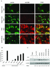
FIG. 1.
FIG. 1.
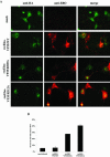
Effects of mutations in the C-terminal domain of VP35 on IRF-3 activation. A) Localization of IRF-3. Vero E6 cells were transfected with HA-tagged IRF-3 and VP35 expression plasmids. Twenty-four hours later, the cells were infected with Sendai virus (+SEV). Six hours postinfection, the cells were fixed and stained with anti-HA (green) or polyclonal anti-Ebola virus (anti-EBO) (red) antibodies. A merged image is shown in the far right panels. Cells transfected with an empty vector (top two rows) show background levels of anti-Ebola virus staining which is distinguishable from wells transfected with VP35 plasmids. B) Percentage of cells with nuclear IRF-3 shown in a graph. For each coverslip, six random fields were chosen and the percentage of cells with nuclear versus cytoplasmic IRF-3 was determined. In total, approximately 250 to 500 cells were counted per sample. The results were averaged, and the standard deviation (error bar) is shown. These data are representative of four independent experiments using various amounts of the VP35 plasmids. C) Lysates made from parallel transfections were subjected to SDS-PAGE and Western blotting for VP35 and actin (loading control). Expression levels of each VP35 protein were measured using a densitometer and normalized to actin expression, which is expressed as a ratio below the panel. α-VP35, anti-VP35 antibody; α-actin, α-actin antibody.
FIG. 2.
FIG. 2.
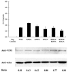
Effects of mutations in the IRF-3 inhibitory domain of VP35 on viral transcription/replication. The 3E-5E CAT minigenome transcription/replication system was used to determine the effects of VP35 mutations on viral transcription. 293T cells were transfected in six-well plates with the 3E-5E minigenome (1 μg) along with expression plasmids for pT7-pol (1 μg), pCEZ-NP (1 μg), pCEZ-VP35 (0.5 μg), pCEZ-VP30 (0.3 μg), and pCEZ-L (1 μg). Forty-eight hours posttransfection, the cells were lysed, and CAT levels were measured by an ELISA. Results shown are averages of wells transfected in duplicate and are representative of three independent experiments. Lysates were subjected to SDS-PAGE and Western blotting to determine VP35 and actin expression levels, and expression ratios were calculated as described in the legend to Fig. 1. Neg Control, negative control.
FIG. 3.
FIG. 3.
Growth of recombinant Ebola viruses and production of RANTES during infection of Vero E6 cells with recombinant Ebola viruses. The growth of the recEBO-VP35/wt, recEBO-VP35/R305A, and recEBO-VP35/R312A viruses was measured in Vero E6 cells at an MOI of 0.02 (A) or 2 (B). Virus titer was determined by standard immunoplaque assay. RANTES was measured in the supernatants from the growth curves by an ELISA. The cells had been infected at an MOI of 0.02 (C) or 2 (D).
FIG. 4.
FIG. 4.
Growth of recEBO-VP35/R312A virus in macrophage and hepatocyte cell lines. The growth of the recEBO-VP35/wt and recEBO-VP35/R312A viruses was measured in U937 macrophages (A) and Huh7 hepatocytes (B) infected at an MOI of 0.02.
FIG. 5.
FIG. 5.
Effect of infection with recombinant Ebola viruses on IRF-3 activation. A) IRF-3 localization. Vero cells were infected with the recombinant Ebola viruses at an MOI of 2 and then transfected with an HA-tagged IRF-3 plasmid. Twenty-four hours after infection, the cells on the coverslips were fixed and stained with anti-HA (green) and anti-Ebola virus (anti-EBO) (red) antibodies. Merged images are shown in the far right panels. B) Percentage of cells containing nuclear IRF-3 is shown in a graph. Approximately 100 to 200 cells were counted per coverslip to determine the ratio of nuclear versus cytoplasmic IRF-3.

References

    1. Baize, S., E. M. Leroy, M. C. Georges-Courbot, M. Capron, J. Lansoud-Soukate, P. Debre, S. P. Fisher-Hoch, J. B. McCormick, and A. J. Georges. 1999. Defective humoral responses and extensive intravascular apoptosis are associated with fatal outcome in Ebola virus-infected patients. Nat. Med. 5:423-426. - PubMed
    1. Barro, M., and J. T. Patton. 2005. Rotavirus nonstructural protein 1 subverts innate immune response by inducing degradation of IFN regulatory factor 3. Proc. Natl. Acad. Sci. USA 102:4114-4119. - PMC - PubMed
    1. Basler, C. F., A. Mikulasova, L. Martinez-Sobrido, J. Paragas, E. Muhlberger, M. Bray, H. D. Klenk, P. Palese, and A. Garcia-Sastre. 2003. The Ebola virus VP35 protein inhibits activation of interferon regulatory factor 3. J. Virol. 77:7945-7956. - PMC - PubMed
    1. Basler, C. F., X. Wang, E. Muhlberger, V. Volchkov, J. Paragas, H. D. Klenk, A. Garcia-Sastre, and P. Palese. 2000. The Ebola virus VP35 protein functions as a type I IFN antagonist. Proc. Natl. Acad. Sci. USA 97:12289-12294. - PMC - PubMed
    1. Becker, S., and E. Muhlberger. 1999. Co- and posttranslational modifications and functions of Marburg virus proteins. Curr. Top. Microbiol. Immunol. 235:23-34. - PubMed

Publication types

MeSH terms