. 2006 Jul;80(13):6517-24.
doi: 10.1128/JVI.02499-05.

Conditionally activated E7 proteins of high-risk and low-risk human papillomaviruses induce S phase in postmitotic, differentiated human keratinocytes

Affiliations

Conditionally activated E7 proteins of high-risk and low-risk human papillomaviruses induce S phase in postmitotic, differentiated human keratinocytes

N Sanjib Banerjee et al. J Virol. 2006 Jul.

Abstract

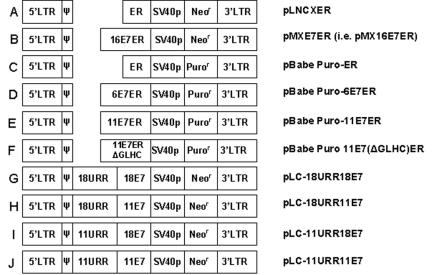
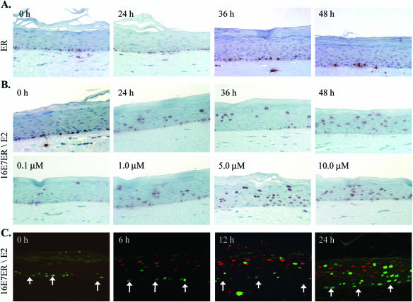
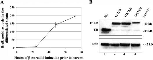
The productive program of human papillomaviruses (HPVs) in epithelia is tightly linked to squamous differentiation. The E7 proteins of high-risk HPV genotypes efficiently inactivate the pRB family of proteins that control the cell cycle, triggering S phase in suprabasal keratinocytes. This ability has until now not been demonstrated for the low-risk HPV-6 or HPV-11 E7 proteins. An inducible system in which HPV-16 E7 is fused to the ligand binding domain of the human estrogen receptor (ER) was described by Smith-McCune et al. (K. Smith-McCune, D. Kalman, C. Robbins, S. Shivakumar, L. Yuschenkoff, and J. M. Bishop, Proc. Natl. Acad. Sci. USA 96:6999-7004, 1999). In the absence of hormone, E7ER is cytoplasmic, and upon addition of 17beta-estradiol, it translocates to the nucleus. Using organotypic epithelial raft cultures developed from primary human keratinocytes, we show that 16E7ER promotes either S-phase reentry or p21cip1 accumulation in differentiated keratinocytes in a stochastic manner as early as 6 h postinduction with 17beta-estradiol. A vector expressing the ER moiety alone had no effect. These observations prove unequivocally that the E7 protein drives S-phase reentry in postmitotic, differentiated keratinocytes rather than preventing S-phase exit while the cells ascend through the epithelium. HPV-11 E7ER and, much less efficiently, HPV-6 E7ER also promoted S-phase reentry by differentiated cells upon exposure to 17beta-estradiol. S-phase induction required the consensus pRB binding motif. We propose that the elevated nuclear levels of the low-risk HPV E7 protein afforded by the inducible system account for the positive results. These observations are entirely consistent with the fact that low-risk HPV genotypes replicate in the differentiated strata in patient specimens, as do the high-risk HPVs.


Figures

FIG. 1.
FIG. 1.
Schematic representation of retroviral vectors used in this study. (A) pLNCXER and (B) pMX16E7ER (35). (C) pBabe Puro-ER expresses the ligand binding domain of human estrogen receptor α from the LTR. (D) pBabe Puro-6E7ER expresses the HPV-6 E7ER fusion from the LTR. (E) pBabe Puro-11E7ER expresses the HPV-11 E7ER fusion from the LTR. (F) pBabe Puro-11E7(ΔGLHC)ER expresses a fusion of ER with HPV-11E7 with critical residues in the pocket protein binding domain from the LTR deleted. (G) pLC-18URR18E7 expresses HPV-18 E7 from the HPV-18 URR. (H) pLC-18URR11E7 expresses HPV-11 E7 from the HPV-18 URR. (I) pLC-11URR18E7 expresses HPV-18 E7 from the HPV-11 URR. (J) pLC-11URR11E7 expresses HPV-11E7 under the control of the HPV-11URR.
FIG. 2.
FIG. 2.
Functional activation of HPV-16 E7ER in PHK raft cultures by E2. The raft cultures were left untreated (0 h) or treated with E2 for the specified durations prior to harvest on day 9. BrdU was added 12 h (A and B) or 6 h (C) prior to harvest. Immunohistochemistry to detect BrdU incorporation (in red) (A and B) was performed on 4-μm sections to reveal S-phase cells. (A) Raft cultures expressing ER after induction with 1 μM E2 for 0 h, 24 h, 36 h, or 48 h. (B) Raft cultures expressing 16E7ER in the absence of E2 or in the presence of 1 μM E2 for 24 h, 36 h, or 48 h (upper images) or after induction for 24 h with 0.1 μM, 1.0 μM, 5.0 μM, or 10 μM E2 (lower images). (C) Double immunofluorescence revealing time-dependent induction of p21cip1 protein stabilization (red) or S-phase reentry, as indicated by BrdU incorporation (green), in separate populations of differentiated cells in the presence of 5 μM of E2 for 0 h, 6 h, 12 h, or 24 h. The arrows point to the basal stratum.
FIG. 3.
FIG. 3.
Ability of the low-risk HPV E7 or E7ER protein to induce unscheduled DNA synthesis in differentiated cells. (A, B, and D) BrdU was added to the raft culture medium 12 h prior to harvest. Four-micrometer sections of raft cultures were probed for BrdU incorporation by immunohistochemistry (in red) to reveal S-phase cells (A) Raft cultures expressing HPV-11 E7 or HPV-18 E7 from either the HPV-11 URR or the HPV-18 URR. (B) Raft cultures expressing HPV-6 E7ER (upper row), HPV-11 E7ER (middle row), or HPV-11 E7(ΔGLHC)ER (lower row) in the absence (0 h) or in the presence of 5 μM of E2 for 24 h, 48 h, or 72 h. (C) Immunofluorescence detection of BrdU (green) in the basal and parabasal layers after a 6-h BrdU pulse followed by 24-h (a and c) or 48-h (b and d) E2 induction. (a and b) Raft cultures of pBabe-Puro vector-transduced PHKs. (c and d) Raft cultures of HPV-11 E7ER-transduced PHKs. Nuclei were labeled with DAPI. (D) S-phase reentry by differentiated cells coincides with nuclear import of E7ER. (a to g) Raft cultures were probed by double indirect immunofluorescence for ER (red) and BrdU (green) incorporation. (a to c) Cultures transduced with pBabe Puro-ER in the absence of E2 (a) or in the presence of 5 μM E2 for 24 h (b) or 48 h (c). (d) A normal untransduced PHK raft culture. (e to h) Cultures transduced with pBabe Puro-11E7ER in the absence of E2 (e) or in the presence of 5 μM E2 for 24 h (f) or 48 h (g). (h) Stabilization of p21cip1 protein (red) and BrdU (green) incorporation at 48 h after induction with 5 μM of E2. Colocalization of p21cip1 and BrdU (yellow) was observed in a small fraction of the differentiated cells. The arrows point to the basal stratum.
FIG. 4.
FIG. 4.
(A) Graphic representation of time-dependent increase in the number of BrdU-positive cells in the differentiated strata following E2-induced nuclear entry of HPV-11 E7ER. The error bars indicate standard deviations. (B) Western blot showing retroviral LTR-driven expression of ER, HPV 6E7ER, HPV-11E7ER, and HPV-16 E7ER after 24 h of E2 induction in submerged cultures of transduced PHK. Equal loading in the same gel was verified by probing for actin.

References

    1. Armstrong, D. J., and A. Roman. 1992. Mutagenesis of human papillomavirus types 6 and 16 E7 open reading frames alters the electrophoretic mobility of the expressed proteins. J. Gen. Virol. 73:1275-1279. - PubMed
    1. Banerjee, N. S., L. T. Chow, and T. R. Broker. 2005. Retrovirus mediated gene transfer to analyze HPV gene regulation and protein functions in “organotypic” raft culture, p. 187-202. In C. Davy and J. Doorbar (ed.), Human papillomaviruses: methods and protocols, methods in molecular medicine. Humana Press, Totowa, N.J. - PubMed
    1. Cheng, S., D.-C. Schmidt-Grimminger, T. Murant, T. R. Broker, and L. T. Chow. 1995. Differentiation-dependent up-regulation of the human papillomavirus E7 gene reactivates cellular DNA replication in suprabasal differentiated keratinocytes. Genes Dev. 9:2335-2349. - PubMed
    1. Chien, W.-M., F. Noya, H. M. Benedict-Hamilton, T. R. Broker, and L. T. Chow. 2002. Alternative fates of keratinocytes transduced by human papillomavirus type 18 E7 during squamous differentiation. J. Virol. 76:2964-2972. - PMC - PubMed
    1. Chien, W.-M., J. N. Parker, D.-C. Schmidt-Grimminger, T. R. Broker, and L. T. Chow. 2000. Casein kinase II phosphorylation of the human papillomavirus-18 E7 protein is critical for promoting S-phase entry. Cell Growth Differ. 11:425-435. - PubMed

Publication types

MeSH terms

LinkOut - more resources