Comparative Study
. 2006 Jul;80(13):6559-67.
doi: 10.1128/JVI.00387-06.

Efficient expression of the adeno-associated virus type 5 p41 capsid gene promoter in 293 cells does not require Rep

Affiliations
Comparative Study

Efficient expression of the adeno-associated virus type 5 p41 capsid gene promoter in 293 cells does not require Rep

Chaoyang Ye et al. J Virol. 2006 Jul.

Abstract

Efficient expression of the adeno-associated virus type 5 (AAV5) P41 capsid gene promoter required adenovirus E1A and/or E1B; however, in contrast to what was observed for expression of the AAV2 capsid gene promoter (P40), neither adenovirus infection nor the large Rep protein was required. Although both the AAV2 and the AAV5 large Rep proteins efficiently bound the (GAGY)(3) Rep-binding element, the AAV5 large Rep protein transactivated transcription of the inducible AAV2 P40 promoter much less well than AAV2 large Rep. Differences in their activation potentials were mapped to the amino-terminal region of the proteins, and the poorly transactivating AAV5 Rep protein could competitively inhibit AAV2 Rep transactivation.


Figures
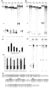
FIG. 1.
FIG. 1.
Transcription profiles of different serotypes of AAV. (A) RNase protection assay, performed as previously described (9, 16), using homologous RP probes (spanning from approximately 86 nt upstream to 54 nt downstream of each P40 or 66 nt upstream and 73 nt downstream of the P41 initiation site) to protect RNA generated in 293 cells following transfection of RepCap constructs of different serotypes of AAV in the absence (−) or presence (+) of adenovirus infection. Bands protected by transcripts from upstream P5 (P7 for AAV5), P19, or P40 (P41 for AAV5) promoters are indicated. Multiple bands for P40 transcripts represent alternative initiation events and some probe chatter; all bands were included in quantifications. (B) This gel shows the same assay performed on RNA generated by transfection of RepStopCap constructs. The AAV2 RepStopCap and AAV5 RepStopCap constructs have premature-termination codons introduced at nt 489 and 480, respectively, in the amino termini of their respective Rep proteins. The AAV1, AAV3, AAV4, and AAV6 RepStopCap constructs contain frameshift mutations in the amino termini of their respective Rep proteins at nt 743, 523, 781, and 729, respectively, which lead to termination of these proteins shortly downstream. (C) Quantification, using a Molecular Imager FX and the Quantity One version 4.2.2 image software (Bio-Rad, Hercules, CA), of RNase protections, a representative of which is shown in panel A. Data from at least three experiments, with standard error bars, are presented as the activation fold of P40 (P41) promoters, comparing transcription levels in the presence or absence of adenovirus. (D) Comprehensive quantification of RNase protections of the type shown in panels A and B. P40 (P41) transcription levels are calculated as the percentages of total transcripts (P5, P19, and P40). Quantifications for at least three individual experiments, including standard error bars, are shown. (E) RNase protection assay, with the same probes as those described for panel A, of RNA generated in HeLa cells following transfection of RepCap and RepStopCap constructs of AAV2 and AAV5. Bands protected by transcripts from upstream P5 (P7 for AAV5) and P19 promoters or P40 (P41 for AAV5) promoters are indicated. (F) Sequence alignment of AAV2 P40 (nt 1690 to 1908) and AAV5 P41 (nt 1726 to 1991) promoters. Consensus nucleotides are shaded. TATA boxes are indicated. SP1 and GGT elements previously reported to mediate AAV2 P40 activation (11) are also indicated.
FIG. 2.
FIG. 2.
AAV2 and AAV5 large Rep proteins have different activation properties. RNase protection assay, performed as previously described (9, 16), using a homologous RNase protection probe spanning either the P40 (nt 1767 to 1906) or the P41 (nt 1846 to 1986) initiation site (designated RP in the diagram at the top of each panel) of RNA generated in 293 cells following transfection of P40VP (nt 1700 to 4492) (14) (A) or P41Cap (nt 1637 to 4448) (B), linked with different upstream Rep-binding sequences (diagramed on the top of each gel). pBluescript SK+ was used as an empty-vector control. AAV2 and AAV5 Rep were provided in trans and driven by HIV promoter as previously described (13, 14). All transfections were followed by adenovirus infection to allow activation. Sequences from AAV2 P5 (nt 146 to 320) (14), AAV5 P7 (nt 168 to 358) (13), the AAV2 ITR (nt 1 to 145) (14), or the AAV5 ITR (nt 1 to 183) (13) were inserted in front of P40VP or P41VP (as shown in the diagram) to create P5P40VP, P7P40VP, V2ITRP40VP, and V5ITRP40VP or P5P41VP, P7P41VP, V2ITRP41VP, and V5ITRP41VP, respectively. The human β-actin probe was used as a loading control. The arrowhead indicates transcripts generated from upstream promoter activities. The asterisk indicates nonspecific bands associated with β-actin protection. The activation fold generated by AAV2 or AAV5 Rep was calculated by setting the empty vector as 1.
FIG. 3.
FIG. 3.
Differences in activation properties of AAV2 and AAV5 Rep reside within the amino terminus. (A) Illustration of the chimeric Rep constructs described in the text. Black bars indicate sequences derived from AAV2 Rep. White bars indicate sequences derived from AAV5 Rep. The numbers in the name of each construct designate exactly where the chimeric borders were made. All fusions were made in frame and sequenced to ensure that they were as predicted. All Rep expression was driven by the HIV promoter, in constructs similar to those previously described (13, 14). The V2 211 (V5) series were based on the V2 211 parent construct. (B, top) diagram of the P5P40 luciferase reporter plasmid used for these assays as described in the text. The AAV2 P5 promoter (nt 146 to 320 from AAV2) was cloned upstream of the P40 (nt 1700 to 1883) luciferase gene, to provide an RBE, but was cloned in an inverted orientation relative to the direction of P40 transcription to prevent P5-generated luciferase activity (Luc Act). pHelper plasmid was cotransfected to provide helper adenovirus gene products. (Bottom) Quantification of wild-type and chimeric Rep transactivation. The activity of each protein is shown with a standard error bar and is the average for duplicate samples from at least three experiments. All values are standardized to β-galactosidase activity generated by a CMV-driven β-galactosidase expression vector used as an internal control.
FIG. 4.
FIG. 4.
(A) AAV2/AAV5 chimeras that have different transactivation properties bind the AAV2 P5 RBE similarly. The P5P40VP reporter plasmid and the RNase protection probe used in this experiment are diagramed. RNase protection assays were performed as previously described (9, 16), using the RP probe described above (as shown in the diagram) on RNA generated in 293 cells following cotransfection of the P5P40VP construct with different Rep chimeras as described in the text and as indicated above each lane of the gel. Protection by the human β-actin probe was used as a control. The arrowhead indicates transcripts generated from upstream promoter activity. The asterisk indicates nonspecific bands associated with β-actin protection. For quantification, all P40 transcription levels were normalized to β-actin levels, and the activity of each fusion protein was reported relative to the P40 activity in response to the pSK+ vehicle (lane 1), which was set to 1. As described in the text, wild-type and chimeric amino-terminal fusions were assayed with and without the VP16 activation domain. (B) AAV5 Rep can competitively inhibit AAV2 Rep-dependent activation of P40. The same luciferase reporter assay as that described in the legend to Fig. 3 was used in this experiment. Activity in response to an HIV-driven AAV2 Rep plasmid was first assayed. The lowest amount of plasmid giving the highest level of activity was determined. These activities (black bar), as well as activity in response to cotransfection of the parent pSK+ (white bar), are shown on the right. In the samples shown on the left, decreasing amounts of AAV2-expressing plasmid (the fraction of which is indicated on the bottom of each pair) were mixed, keeping the final DNA concentration the same, with either empty vector (white bars) or a plasmid expressing AAV5 Rep from an HIV promoter (black bars). At each amount of AAV2 Rep-expressing plasmid used, the cotransfected excess AAV5 Rep-expressing plasmid reduced AAV2 Rep-dependent activation more than did equal amounts of cotransfected vehicle. Each experiment represents the average values for duplicate samples from three individual experiments (error bars shown), each normalized to β-galactosidase activity expressed from a cotransfected expression vector in which β-galactosidase was driven by a CMV promoter. Luc Act, luciferase activity.

Similar articles

Cited by

References

    1. Cathomen, T., D. Collete, and M. D. Weitzman. 2000. A chimeric protein containing the N terminus of the adeno-associated virus Rep protein recognizes its target site in an in vivo assay. J. Virol. 74:2372-2382. - PMC - PubMed
    1. Chiorini, J. A., S. Afione, and R. M. Kotin. 1999. Adeno-associated virus (AAV) type 5 Rep protein cleaves a unique terminal resolution site compared with other AAV serotypes. J. Virol. 73:4293-4298. - PMC - PubMed
    1. Chiorini, J. A., L. Yang, Y. Liu, B. Safer, and R. M. Kotin. 1997. Cloning of adeno-associated virus type 4 (AAV4) and generation of recombinant AAV4 particles. J. Virol. 71:6823-6833. - PMC - PubMed
    1. Hickman, A. B., D. R. Ronning, R. M. Kotin, and F. Dyda. 2002. Structural unity among viral origin binding proteins: crystal structure of the nuclease domain of adeno-associated virus Rep. Mol. Cell 10:327-337. - PubMed
    1. Hickman, A. B., D. R. Ronning, Z. N. Perez, R. M. Kotin, and F. Dyda. 2004. The nuclease domain of adeno-associated virus rep coordinates replication initiation using two distinct DNA recognition interfaces. Mol. Cell 13:403-414. - PubMed

Publication types

MeSH terms

Substances

LinkOut - more resources