. 2006 Jul;80(13):6691-6.
doi: 10.1128/JVI.00057-06.

Identification of a dynein interacting domain in the papillomavirus minor capsid protein l2

Affiliations

Identification of a dynein interacting domain in the papillomavirus minor capsid protein l2

Luise Florin et al. J Virol. 2006 Jul.

Abstract

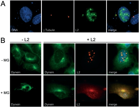
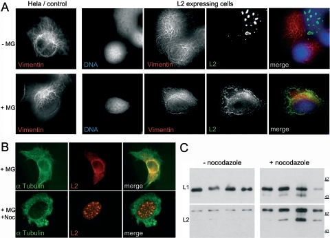
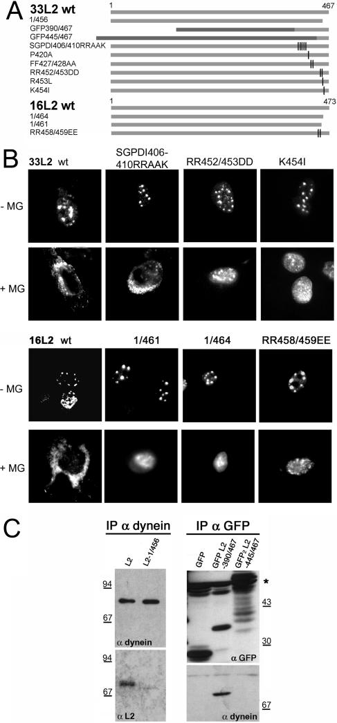
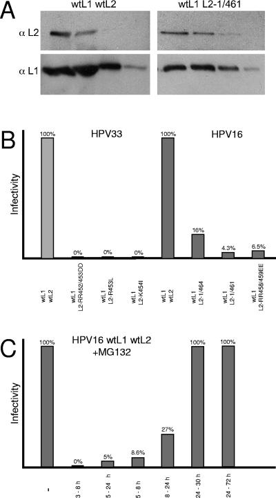
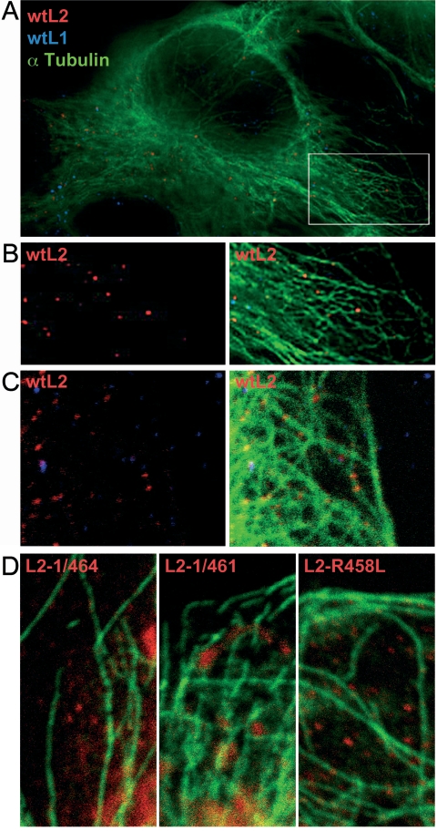
Papillomaviruses enter cells via endocytosis (H. C. Selinka et al., Virology 299:279-287, 2002). After egress from endosomes, the minor capsid protein L2 accompanies the viral DNA to the nucleus and subsequently to the subnuclear promyelocytic leukemia protein bodies (P. M. Day et al., Proc. Natl. Acad. Sci. USA 101:14252-14257, 2004), suggesting that this protein may be involved in the intracytoplasmic transport of the viral genome. We now demonstrate that the L2 protein is able to interact with the microtubule network via the motor protein dynein. L2 protein was found attached to microtubules after uncoating of incoming human papillomavirus pseudovirions. Based on immunofluorescence and coimmunoprecipitation analyses, the L2 region interacting with dynein is mapped to the C-terminal 40 amino acids. Mutations within this region abrogating the L2/dynein interaction strongly reduce the infectivity of pseudoviruses, indicating that this interaction mediates the minus-end-directed transport of the viral genome along microtubules towards the nucleus.


Figures

FIG. 1.
FIG. 1.
Coimmunofluorescence of L2 and γ-tubulin (A) or dynein (B), respectively. HPV33 L2 was expressed in HeLa cells using recombinant vaccinia virus vac33L2 (29) in the absence (A, B) or presence of 10 μM MG132 (B). MG132 was added 3 h postinfection. Cells were fixed and stained 6 h later using a mouse monoclonal antibody against the dynein intermediate chain (Abcam) and L2-specific polyclonal rabbit antiserum K28 (31).
FIG. 2.
FIG. 2.
Analysis of L2 function in the presence of MG132. HPV16 L2-expressing HeLa cells were stained for vimentin (mouse monoclonal antibody; Sigma) and L2 (K18) (31) and observed by fluorescence microscopy using 3D deconvolution (A). HeLa cells were treated as described in the Fig. 1 legend in the presence or absence of nocodazole and stained with L2- and α-tubulin-specific (Sigma) antibodies (B). VLPs obtained from HeLa cells treated with MG132 in the absence or presence of nocodazole were subjected to sucrose gradient centrifugation. Peak fractions were analyzed for L2 incorporation by Western blotting using L1- and L2-specific antibodies L1-7 and L2-1, respectively (23, 32) (C).
FIG. 3.
FIG. 3.
Mapping the L2/dynein interaction domain. (A)Schematic representation of HPV33 and HPV16 L2 mutants. Light-gray bars symbolize L2, dark-gray bars symbolize GFP. The 445/467 sequence of 33L2 is fused to dimeric GFP (14). (B) Immunofluorescent detection of L2 deletion and point mutants expressed in HuTK-143B cells in the absence or presence of MG132. (C) Coimmunoprecipitation analysis of L2, L2 mutants, and L2 fusion proteins with dynein, followed by Western blotting using dynein-, L2-, and GFP-specific antibodies. wt, wild type.
FIG. 4.
FIG. 4.
Infectivity of mutant HPV33 and HPV16 pseudovirions. Peak fractions of Optiprep gradients loaded with wild-type (wt) and mutant pseudovirions, respectively, were analyzed for the presence of L1 and L2 by Western blotting (A). Cells were infected with equal amounts of wild-type or mutant pseudovirions. Infectious events were monitored 72 h postinfection by counting cells exhibiting green nuclear fluorescence (B). Cells were infected with wild-type HPV16 pseudovirions in the absence or presence of 10 μM MG132 at the time periods indicated (C).
FIG. 5.
FIG. 5.
(A) Triple staining of HuTK-143B cells infected with HPV16 pseudovirions using 3D deconvolution. (B) Enlarged section. (C) Representative section of a confocal microscopic analysis. (D) Representative section of a confocal microscopic analysis of cells infected with mutant HPV16 PsV harboring 16L2-1/464, -1/461, or -R458L. Sections were processed for immunofluorescence 20 h postinfection. wt, wild type.

References

    1. Becker, K. A., L. Florin, C. Sapp, G. G. Maul, and M. Sapp. 2004. Nuclear localization but not PML protein is required for incorporation of the papillomavirus minor capsid protein L2 into virus-like particles. J. Virol. 78:1121-1128. - PMC - PubMed
    1. Becker, K. A., L. Florin, C. Sapp, and M. Sapp. 2003. Dissection of human papillomavirus type 33 L2 domains involved in nuclear domain (ND) 10 homing and reorganization. Virology 314:161-167. - PubMed
    1. Buck, C. B., D. V. Pastrana, D. R. Lowy, and J. T. Schiller. 2004. Efficient intracellular assembly of papillomaviral vectors. J. Virol. 78:751-757. - PMC - PubMed
    1. Darshan, M. S., J. Lucchi, E. Harding, and J. Moroianu. 2004. The L2 minor capsid protein of human papillomavirus type 16 interacts with a network of nuclear import receptors. J. Virol. 78:12179-12188. - PMC - PubMed
    1. Day, P. M., C. C. Baker, D. R. Lowy, and J. T. Schiller. 2004. Establishment of papillomavirus infection is enhanced by promyelocytic leukemia protein (PML) expression. Proc. Natl. Acad. Sci. USA 101:14252-14257. - PMC - PubMed

Publication types

MeSH terms

Substances

LinkOut - more resources