. 2006 Aug 15;575(Pt 1):49-67.
doi: 10.1113/jphysiol.2006.114074. Epub 2006 Jun 15.

Angiotensin II regulates neuronal excitability via phosphatidylinositol 4,5-bisphosphate-dependent modulation of Kv7 (M-type) K+ channels

Affiliations

Angiotensin II regulates neuronal excitability via phosphatidylinositol 4,5-bisphosphate-dependent modulation of Kv7 (M-type) K+ channels

Oleg Zaika et al. J Physiol. .

Abstract

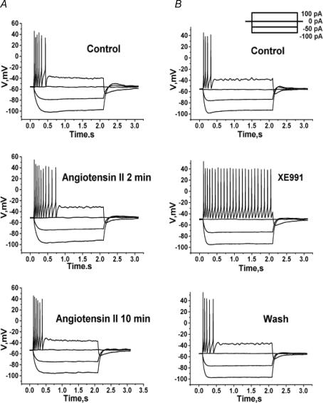
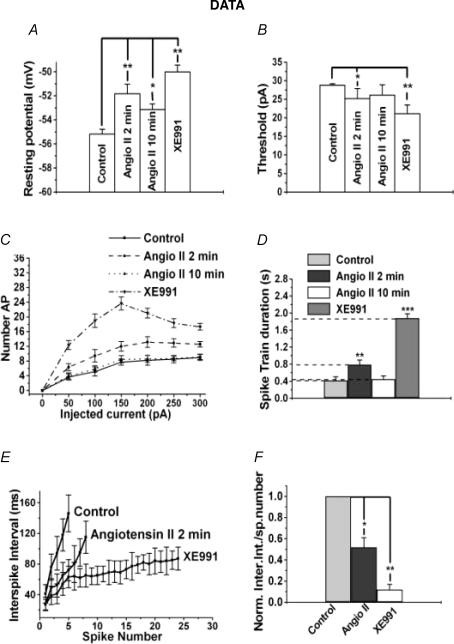
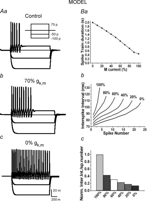
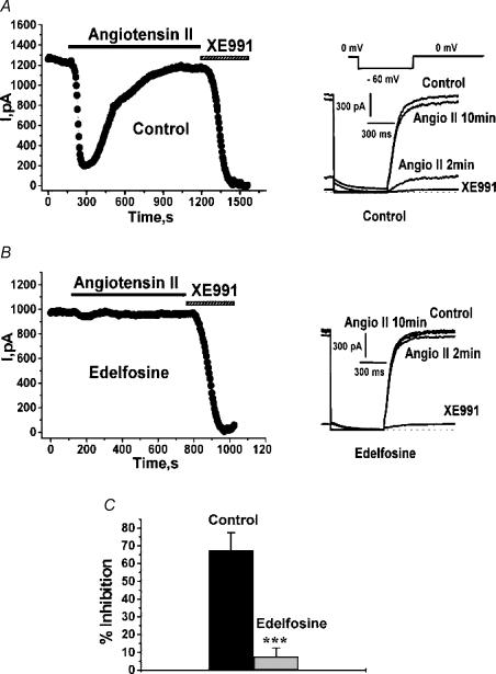
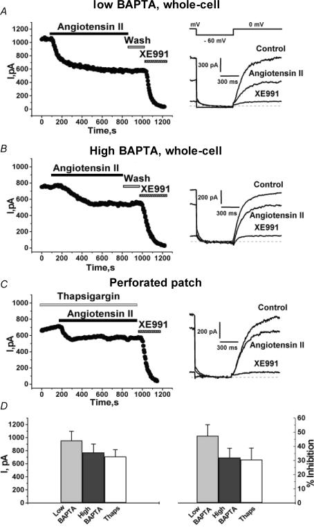
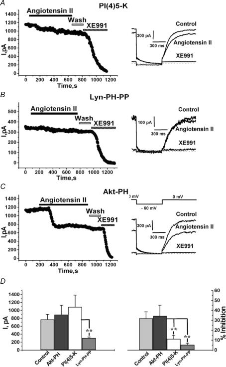
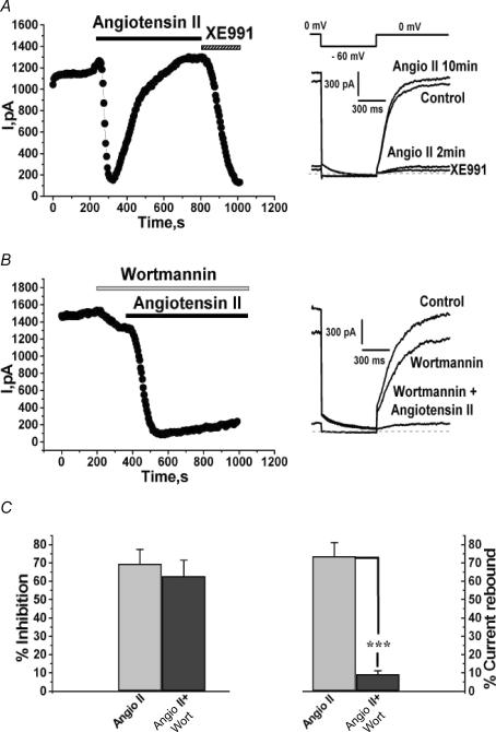
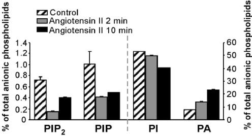
Voltage-gated Kv7 (KCNQ) channels underlie important K+ currents in many different types of cells, including the neuronal M current, which is thought to be modulated by muscarinic stimulation via depletion of membrane phosphatidylinositol 4,5-bisphosphate (PIP2). We studied the role of modulation by angiotensin II (angioII) of M current in controlling discharge properties of superior cervical ganglion (SCG) sympathetic neurons and the mechanism of action of angioII on cloned Kv7 channels in a heterologous expression system. In SCG neurons, which endogenously express angioII AT1 receptors, application of angioII for 2 min produced an increase in neuronal excitability and a decrease in spike-frequency adaptation that partially returned to control values after 10 min of angioII exposure. The increase in excitability could be simulated in a computational model by varying only the amount of M current. Using Chinese hamster ovary (CHO) cells expressing cloned Kv7.2 + 7.3 heteromultimers and AT1 receptors studied under perforated patch clamp, angioII induced a strong suppression of the Kv7.2/7.3 current that returned to near baseline within 10 min of stimulation. The suppression was blocked by the phospholipase C inhibitor edelfosine. Under whole-cell clamp, angioII moderately suppressed the Kv7.2/7.3 current whether or not intracellular Ca2+ was clamped or Ca2+ stores depleted. Co-expression of PI(4)5-kinase in these cells sharply reduced angioII inhibition, but did not augment current amplitudes, whereas co-expression of a PIP2 5'-phosphatase sharply reduced current amplitudes, and also blunted the inhibition. The rebound of the current seen in perforated-patch recordings was blocked by the PI4-kinase inhibitor, wortmannin (50 microM), suggesting that PIP2 re-synthesis is required for current recovery. High-performance liquid chromatographic analysis of anionic phospholipids in CHO cells stably expressing AT1 receptors revealed that PIP2 and phosphatidylinositol 4-phosphate levels are to be strongly depleted after 2 min of stimulation with angioII, with a partial rebound after 10 min. The results of this study establish how angioII modulates M channels, which in turn affects the integrative properties of SCG neurons.


Figures

Figure 1
Figure 1. AngioII increases the somatic excitability of SCG neurons
Cultured SCG neurons were studied under perforated-patch current clamp, and voltage responses were recorded from a set of current pulses (inset). A, voltage sweeps are shown before (top), after 2 min (middle) or after 10 min (bottom) bath application of angioII (500 nm). B, voltage sweeps are shown before (top), during bath application of XE991 (10 μm) (middle) and after washout (bottom).
Figure 2
Figure 2. Analysis of effects of angioII on SCG discharge properties
Bars show summarized measurements for neurons of the resting potential (A), the threshold current for AP generation (B), or the spike-train duration (D) in control, after 2 min or 10 min bath application of angioII, or after application of XE991. C, shows the number of APs evoked during the current step versus current amplitude for neurons in the above cases. E, shows the instantaneous interspike interval versus spike number relation for control, after 2 min application of angioII and in the presence of XE991. F, the data in E are shown normalized, calculated as the difference between the initial and final interspike interval of the spike train divided by the number of spikes.
Figure 3
Figure 3. A computational model of an SCG neuron simulates the effect of M channel closure on firing properties
The data in Figs 1 and 2 were used to constrain the parameters of a computational model of an SCG neuron (see Methods) that simulated the effects on firing of various degrees of M current closure. AaAc, show simulated voltage responses to the set of current injections shown in the inset in the cases of undiminished M current (control), 30% M current blockade (70% gK,M) or 100% M current blockade (0% gK,M). Ba, shows the simulated relationship between spike-train duration and percentage gK,M values. Bb, shows the simulated curves of interspike interval versus spike number at different percentage gK,M values. Bc, the slopes of the curves in Bb are normalized, as in Fig. 2F.
Figure 4
Figure 4. Suppression of Kv7.2/7.3 currents in CHO cells by angioII requires PLC
CHO cells were transiently transfected with Kv7.2 + Kv7.3 subunits and AT1 receptors and studied under perforated-patch voltage clamp. Cells were incubated for 30 min with vehicle alone (A) or edelfosine (10 μm) (B) before starting the experiment. Plotted are current amplitudes of currents elicited by the pulse protocol shown in the inset. AngioII (500 nm) or XE991 (50 μm) were bath-applied during the period indicated by the bars, and representative current traces are shown on the right. C, bars show summarized current inhibitions for control cells and for those incubated with edelfosine.
Figure 5
Figure 5. Stimulation of angioII AT1 receptors suppresses Kv7.2/7.3 channels independently of [Ca2+]i
CHO cells were transiently transfected with Kv7.2 + 7.3 subunits and AT1 receptors (A and B), or CHO cells stably expressing AT1 receptors were transiently transfected with Kv7.2 + 7.3 subunits (C). The whole-cell configuration was used with pipettes containing either the normal (low BAPTA) (A) or ‘cocktail’ (see Methods) (B) pipette solution, or the perforated-patch mode was used (C). AngioII (500 nm), XE991 (50 μm) and thapsigargin (2 μm) were bath applied during the periods shown by the bars. Current traces in control, and after application or angioII or XE991 are shown on the right, using the indicated voltage-pulse protocol. D, shows the summarized current amplitudes (left) or percentage inhibitions (right) for these three groups of cells.
Figure 6
Figure 6. Over-expression of a PIP2 kinase or phosphatase affects the current amplitudes and modulation by angioII of Kv7.2/7.3 channels
CHO cells were transiently transfected with Kv7.2 + 7.3 channels and AT1 receptors, together with the PI(4)5-kinase (A), Lyn-PH-PP (B) or Akt-PH (C) constructs. AngioII (500 nm) or XE991 (50 μm) were bath applied during the periods shown by the bars. Current traces in control, and after application or angioII or XE991 are shown on the right, using the indicated voltage-pulse protocol. D, shows the summarized current amplitudes (left) or percentage inhibitions (right) for these three groups of cells. **P < 0.01.
Figure 7
Figure 7. Current rebound from AT1 receptor desensitization requires PIP2 re-synthesis
A and B, CHO cells stably expressing AT1 receptors were transiently transfected with Kv7.2 + 7.3 subunits and studied under perforated patch. AngioII (500 nm), XE991 (50 μm) and wortmannin (30 μm) were bath applied during the periods shown by the bars. Current traces at the indicated times are shown on the right, using the voltage-pulse protocol shown above. C, shows the percentage inhibitions (left) or the percentage current rebound after the presumed desensitization of the AT1 receptors (right) for these two groups of cells. ***P < 0.001.
Figure 8
Figure 8. Stimulation of cloned AT1 receptors depletes PIP2
CHO cells stably expressing AT1 receptors were stimulated with angioII (2 μm) for 2 or 10 min. Bars show the fractional abundance of anionic phospholipids at the indicated times, expressed as a percentage of the total pool. The left ordinate is for the PIP2 and PIP data, and the right ordinate is for the PI and PA data.

References

    1. Aileru AA, Logan E, Callahan M, Ferrario CM, Ganten D, Diz DI. Alterations in sympathetic ganglionic transmission in response to angiotensin II in (mRen2)27 transgenic rats. Hypertension. 2004;43:270–275. - PubMed
    1. Akasu T, Hirai K, Koketsu K. Modulatory actions of ATP on membrane potentials of bullfrog sympathetic ganglion cells. Brain Res. 1983;258:313–317. - PubMed
    1. Alkadhi KA, Alzoubi KH, Aleisa AM. Plasticity of synaptic transmission in autonomic ganglia. Prog Neurobiol. 2005;75:83–108. - PubMed
    1. Beech DJ, Bernheim L, Mathie A, Hille B. Intracellular Ca2+ buffers disrupt muscarinic suppression of Ca2+ current and M current in rat sympathetic neurons. Proc Natl Acad Sci U S A. 1991;88:652–656. - PMC - PubMed
    1. Bender K, Wellner-Kienitz MC, Pott L. Transfection of a phosphatidyl-4-phosphate 5-kinase gene into rat atrial myocytes removes inhibition of GIRK current by endothelin and alpha-adrenergic agonists. FEBS Lett. 2002;529:356–360. - PubMed

Publication types

MeSH terms