. 2006 Jun 27;103(26):9832-7.
doi: 10.1073/pnas.0602398103. Epub 2006 Jun 15.

Mechanism of action of a flavin-containing monooxygenase

Affiliations

Mechanism of action of a flavin-containing monooxygenase

Subramaniam Eswaramoorthy et al. Proc Natl Acad Sci U S A. .

Erratum in

  • Proc Natl Acad Sci U S A. 2007 Sep 4;104(36):14543

Abstract

Elimination of nonnutritional and insoluble compounds is a critical task for any living organism. Flavin-containing monooxygenases (FMOs) attach an oxygen atom to the insoluble nucleophilic compounds to increase solubility and thereby increase excretion. Here we analyze the functional mechanism of FMO from Schizosaccharomyces pombe using the crystal structures of the wild type and protein-cofactor and protein-substrate complexes. The structure of the wild-type FMO revealed that the prosthetic group FAD is an integral part of the protein. FMO needs NADPH as a cofactor in addition to the prosthetic group for its catalytic activity. Structures of the protein-cofactor and protein-substrate complexes provide insights into mechanism of action. We propose that FMOs exist in the cell as a complex with a reduced form of the prosthetic group and NADPH cofactor, readying them to act on substrates. The 4alpha-hydroperoxyflavin form of the prosthetic group represents a transient intermediate of the monooxygenation process. The oxygenated and reduced forms of the prosthetic group help stabilize interactions with cofactor and substrate alternately to permit continuous enzyme turnover.


Conflict of interest statement

Conflict of interest statement: No conflicts declared.

Figures

Fig. 1.
Fig. 1.
FMO activity assay. The OD reading at 412 nm due to the DTNB formation in the presence of the substrate methimazole is plotted against time. Dots represent experimental points, and the smoothened curve is given as a solid line. Oxygenated substrate formation is confirmed by this experiment.
Fig. 2.
Fig. 2.
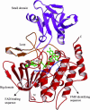
Ribbon representation of the protein and ball-and-stick model of FAD. The strand–turn–helix motifs and the loop interlinking the two domains are labeled. FAD is in the large domain and has no interaction with the small domain.
Fig. 3.
Fig. 3.
Protein–cofactor and protein–substrate interactions. (a) Electrostatic potential of the large domain of FMO. The insertion domain was excluded to clarify the view of the cavity formed along the large domain. FAD is depicted as a stick model. This cavity accommodates the prosthetic group, with adenine completely buried in the protein, and the flavin is more exposed to the solvent. (b) Hydrogen bonding interactions of the prosthetic group. FAD is represented as a ball-and-stick model, and the protein residues are shown as sticks. Water molecules are shown as red spheres. (c) Electrostatic potential of the insertion domain of FMO. The NADPH cofactor is shown as a stick model. NADPH is bound to the protein in a shallow cavity. (d) Hydrogen bonding interactions of the cofactor. NADPH is rendered as a ball-and-stick model, and the protein residues are shown as sticks. Water molecules are shown as red spheres. (e) Stereo diagram of methimazole and the isoalloxazine ring stacking along with a nearby water molecule and Asn-91. The hydrogen bonding interactions are shown by dashed lines. Long dashed lines represent the possible interaction routes involved in the oxygen transfer to the substrate. Two water molecules bonded with a solid line represent the molecular oxygen.
Fig. 4.
Fig. 4.
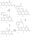
Schematic representation of the functional mechanism of FMO. Only the relevant parts required to explain the function, isoalloxazine, nicotinamide, and methimazole, are shown. Step 1 is seen in the wild type, step 3 is seen in the protein–cofactor complex, and step 5 is seen in the protein–methimazole complex structures.

References

    1. Dixit A., Roche T. E. Arch. Biochem. Biophys. 1984;233:50–63. - PubMed
    1. Poulsen L. L., Ziegler D. J. Biol. Chem. 1979;254:6449–6455. - PubMed
    1. Fraaije M. W., Kamerbeek N. M., van-Berkel W. J. J., Janssen D. B. FEBS Lett. 2002;518:43–47. - PubMed
    1. Ziegler D. M. Annu. Rev. Pharmocol. Toxicol. 1993;33:179–199. - PubMed
    1. Cashman J. R. Curr. Drug Metab. 2000;1:181–191. - PubMed

Publication types

MeSH terms

LinkOut - more resources