. 2006 Oct 15;108(8):2836-45.
doi: 10.1182/blood-2006-04-016394. Epub 2006 Jun 15.

MBD2 is a critical component of a methyl cytosine-binding protein complex isolated from primary erythroid cells

Affiliations

MBD2 is a critical component of a methyl cytosine-binding protein complex isolated from primary erythroid cells

Evan P Kransdorf et al. Blood. .

Abstract

The chicken embryonic beta-type globin gene, rho, is a member of a small group of vertebrate genes whose developmentally regulated expression is mediated by DNA methylation. Previously, we have shown that a methyl cytosine-binding complex binds to the methylated rho-globin gene in vitro. We have now chromatographically purified and characterized this complex from adult chicken primary erythroid cells. Four components of the MeCP1 transcriptional repression complex were identified: MBD2, RBAP48, HDAC2, and MTA1. These 4 proteins, as well as the zinc-finger protein p66 and the chromatin remodeling factor Mi2, were found to coelute by gel-filtration analysis and pull-down assays. We conclude that these 6 proteins are components of the MeCPC. In adult erythrocytes, significant enrichment for MBD2 is seen at the inactive rho-globin gene by chromatin immunoprecipitation assay, whereas no enrichment is observed at the active beta(A)-globin gene, demonstrating MBD2 binds to the methylated and transcriptionally silent rho-globin gene in vivo. Knock-down of MBD2 resulted in up-regulation of a methylated rho-gene construct in mouse erythroleukemic (MEL)-rho cells. These results represent the first purification of a MeCP1-like complex from a primary cell source and provide support for a role for MBD2 in developmental gene regulation.


Figures

Figure 1.
Figure 1.
The chicken homolog of MBD2 is a bona fide methyl-CpG-binding protein and is a component of the MeCPC. (A) DNA sequences of the 50-bp probes used for EMSA with cMBD2. The sequence is derived from the first exon of the ρ-globin gene. The unmodified sequence contains 1 CpG (a1CpG). The sequence was modified to contain no CpGs (a0CpG), 2 CpGs (a2CpG), and 3 CpGs (a3CpG). All CpGs are indicated in bold. Bases that have been mutated compared with the wild-type ρ-globin gene are underlined. (B) cMBD2 is a bona fide MCBP in vitro. The probes a0CpG, a1CpG, a2CpG, and a3CpG were methylated and incubated with recombinant cMBD2. The binding reactions were subjected to EMSA. cMBD2 forms a complex (indicated by the arrow) with the probes containing methyl-CpGs. (C) Anti-cMBD2 antibodies supershift the MeCPC. Inclusion of V2 anti-cMBD2 IgG, but not control IgG, retarded the mobility of the MeCPC complex (supershift) during EMSA.
Figure 2.
Figure 2.
Identification of components of the MeCPC purified from primary erythroid cells. (A) Chromatographic scheme (strategy I) used to purify the MeCPC from primary erythroid cells. The fractions eluted from each column were assayed for MeCPC activity by EMSA using the M-ρ248 probe. (B) EMSA on the eluted fractions from the final column of MeCPC purification strategy I (Heparin Sepharose HP) using the M-ρ248 probe. The MeCPC elutes in fractions 32 through 36. After 4 column chromatography steps the complex remains intact. (C, left) Sypro Ruby-stained protein gel containing 15 μg purified MeCPC. The identity of the bands was determined by matrix-assisted laser desorption/ionization time-of-flight (MALDI-TOF) mass spectrometry followed by peptide mass fingerprint data analysis. The molecular mass of markers in kDa is indicated. (Right) Identities of the bands in the gel. Proteins are grouped into columns based on the complex the protein has previously been associated with (MeCP1, eIF3, or Other). Four components of the MeCP1 complex were identified in the purified MeCPC sample: MBD2, RBAP48, HDAC2, and MTA1. Four components of the eIF3 complex as well as an associated protein were identified: eIF3i, eIF3h, eIF3e, eIF3d, and HSPC021. Five additional proteins were identified: β-actin, MENT, p50unk, BAF60b, and FMIP. The identity of the 2 smallest proteins in the gel could not be established.
Figure 3.
Figure 3.
Putative components of the MeCPC copurify from a Superose 6 gel-filtration column. (A) The second chromatographic scheme (strategy II) was used to purify the MeCPC from primary erythroid cells. The fractions eluted from each column were assayed for MeCPC activity by EMSA using the M-ρ248 probe. (B) EMSA on the high molecular weight fractions from the final column of MeCPC purification strategy II (Superose 6) using the methylated M-ρ248 probe. The MeCPC elutes primarily in fractions 8 through 15 in a molecular weight range of 670 to 2000 kDa. The position of the MeCPC is indicated as “M,” the slower-migrating supercomplex as “Sup,” and the faster-migrating subcomplex as “Sub.” Although the exact composition of the supercomplex is unknown, we hypothesize that the subcomplex results from cleavage of MeCPC component proteins during biochemical purification. (C) EMSA on the high molecular weight fractions from the final column of MeCPC purification strategy II (Superose 6) using the unmethylated ρ248 probe. Both the MeCPC and the supercomplex fail to retard the mobility of an unmethylated probe. In contrast, the subcomplex is able to retard the ρ248 probe (fractions 18 and 19). (D) Western blot analysis of the high molecular weight fractions from the Superose 6 gel-filtration column. The antibody used for each blot is indicated at the right. The putative MeCPC components MBD2, RBAP48, HDAC2, p66, MTA1, and Mi2 copurify from the column. In addition, the erythroid-specific heterochromatin protein MENT also copurifies with the MeCPC factors.
Figure 4.
Figure 4.
cMBD2 affinity copurifies the other components of the MeCP1 complex. (A) Western blot analysis of cMBD2 and streptavidin expression in 6C2 cells containing the BAP-cMBD2 construct with or without the biotin ligase BirA. Equal expression of cMBD2 is seen in BirA- and BirA+ BAP-cMBD2 6C2 cells, whereas biotinylation of cMBD2 only occurs in the BirA+ cells. (B) Western blot analysis of the eluate from cMBD2 pull-down experiments in BAP-cMBD2 6C2 cells. Abundant biotinylated cMBD2 as well as the major components of the MeCP1 complex are seen in the eluate from pull-down assays performed using BirA+ BAP-cMBD2 6C2 cells as the input. In contrast, no biotinylated cMBD2 or any MeCP1 components are seen in the eluate from pull-down assays performed using BirA- BAP-cMBD2 6C2 cells as the input. (C) Sypro Ruby-stained protein gel of the eluate from cMBD2 pull-down experiments in BAP-cMBD2 6C2 cells. Interestingly, despite equal amounts of input protein, less protein elutes from streptavidin beads bound to BirA+ BAP-cMBD2 6C2 cell nuclear extract. The identities of bands in the gel were determined by tandem-mass spectrometry on excised and trypsin-digested bands. The identities of the indicated bands are listed on the right. At least 2 experimentally derived peptides were required for protein identification.
Figure 5.
Figure 5.
MBD2 occupancy inversely correlates with transcription and histone H3-lysine 4-trimethylation (H3-K4-Me3) at the ρ- and βA-globin genes. (A) Enrichment for MBD2, H3-K4-Me3, and IgG at the ρ-globin gene in 5-day (formula image
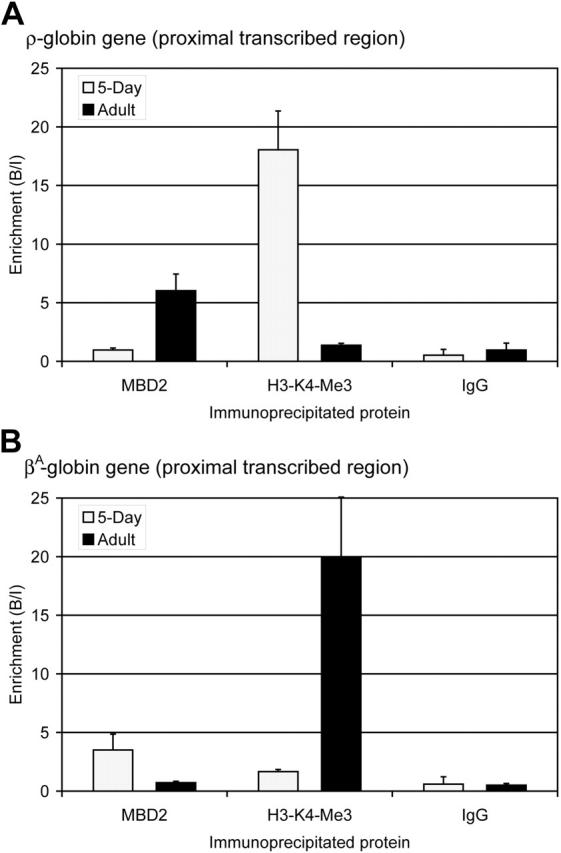
) and adult (▪) erythrocytes as determined by ChIP assay. The data show that MBD2 is depleted from the transcriptionally active ρ-globin gene in 5-day erythrocytes but enriched at the transcriptionally inactive and methylated ρ-globin gene in adult erythrocytes. In contrast, H3-K4-Me3 is enriched at the transcriptionally active ρ-globin gene and depleted at the transcriptionally inactive gene. No enrichment was seen using anti-rabbit IgG in either 5-day or adult erythrocytes, verifying the interaction of these specific proteins with the ρ-globin gene. The data represent the average of 3 independent experiments, with the SD indicated by the bar. (B) Enrichment for MBD2, H3-K4-Me3, and IgG at the βA-globin gene in 5-day (formula image) and adult (▪) erythrocytes as determined by ChIP assay. The data show that MBD2 is enriched at the transcriptionally inactive and methylated βA-globin gene in 5-day erythrocytes but depleted from the transcriptionally active βA-globin gene in adult erythrocytes. Once again, H3-K4-Me3 is depleted from the transcriptionally inactive βA-globin gene and enriched at the transcriptionally active gene. No enrichment was seen using anti-rabbit IgG in either 5-day or adult erythrocytes, verifying the interaction of these specific proteins with the βA-globin gene. The data represent the average of 3 independent experiments, with the SD indicated by the bar.
Figure 6.
Figure 6.
MBD2 is a critical component of the MeCPC in primary mouse splenocytes and MEL-ρ cells. (A) Graphic depiction of the ρ-globin mini-locus introduced into MEL cells. The locus contains (1) a 4.5-kb ρ-globin genomic sequence, (2) a 4-kb chicken LCR enhancer element (HSS2 and HSS3), and (3) 5′ and 3′ cHS4 insulator elements that surround the gene and enhancer. A 2.5-kb fragment of the ρ-globin genomic sequence extending from 248 bp upstream to 2.2 kb downstream of the cap site was excised, in vitro methylated, and religated prior to transfection into MEL cells. In this way the ρ-globin gene is methylated at the same sites as the endogenous gene in chicken adult erythroid cells. (B) EMSA performed with 20 μg nuclear extract from primary mouse splenocytes. Extracts derived from spleens of MBD2+/+ mice form the MeCPC and can be supershifted by the addition of anti-mMBD2 IgG but not control IgG. In contrast, extracts derived from the spleens of MBD2-/- mice do not form a complex on the M-ρ248 probe. (C) RNase protection assay analyzing expression of ρ-globin, mMBD2, and 18S RNAs in MEL-ρ cells treated with shRNAs targeting mMBD2. Significant knock-down of mMBD2 expression is seen in MEL-ρ cells containing shRNA-expressing plasmids that target mMBD2, as compared with control cells (lanes 3 and 4 as compared with lanes 1 and 2). No ρ-globin expression is seen in MEL-ρ cells with wild-type MBD2 expression (lane 2). In contrast, robust ρ-globin expression is seen in MEL-ρ cells in which mMBD2 expression has been knocked down by shRNA (lane 4). Loading of RNA for all MEL samples was equal, as shown by equal amounts of 18S RNA present in the samples. This datum indicates that MBD2 is functionally required for full transcriptional silencing of the methylated ρ-globin gene. (D) Slot blots performed with genomic DNA from wild-type MEL or MEL-ρ cells that contain a stably integrated ρ-globin mini-locus. Genomic DNA from MEL-ρ cells contains ρ-globin gene sequences, whereas wild-type MEL cells display only background hybridization. A 2-fold difference in copy number is observed between mMBD2+ MEL-ρ and mMBD2- knock-down MEL-ρ cells. In contrast, a similar amount of endogenous mouse GAPDH gene sequences is seen in all 3 samples, demonstrating equal DNA loading. Chicken genomic DNA was used as a positive control for the ρ-globin probe.
Figure 7.
Figure 7.
Model for the developmental regulation of ρ-globin transcription in chicken erythrocytes. (A) In embryonic day 4 primitive chicken erythrocytes, robust expression of the ρ-globin gene is seen. The presence of positively-acting trans factors such as GATA1 recruits RNA polymerase (RNAPII) and drives high levels of transcription from the unmethylated ρ-globin gene. Histones throughout the gene exhibit high levels of transcriptionally active modifications, such as trimethylation of H3-K4 as well as H3 and H4 acetylation. (B) No transcription of the ρ-globin gene is seen in adult definitive chicken erythrocytes. In these cells there is dense methylation at the promoter, ρ-PTR, and downstream regions of the gene. In this work we have shown that cMBD2 binds to the methylated ρ-globin gene in adult cells. Because cMBD2 can affinity copurify the other components of the MeCPC complex in vivo, it is likely that MBD2 recruits these components to the methylated ρ-globin gene. Indeed, we show that MBD2 and MTA2 occupy the methylated gene in MEL-ρ cells. The complex maintains transcriptional inactivity by remodeling chromatin into a nonpermissive configuration. Coincident with this loss of transcriptional activity is the loss of trimethylation of H3-K4.

References

    1. McGhee JD, Ginder GD. Specific DNA methylation sites in the vicinity of the chicken beta-globin genes. Nature. 1979;280: 419-420. - PubMed
    1. Shen CK, Maniatis T. Tissue-specific DNA methylation in a cluster of rabbit beta-like globin genes. Proc Natl Acad Sci U S A. 1980;77: 6634-6638. - PMC - PubMed
    1. van der Ploeg LH, Flavell RA. DNA methylation in the human gamma delta beta-globin locus in erythroid and nonerythroid tissues. Cell. 1980;19: 947-958. - PubMed
    1. Groudine M, Weintraub H. Activation of globin genes during chicken development. Cell. 1981; 24: 393-401. - PubMed
    1. Herman JG, Baylin SB. Gene silencing in cancer in association with promoter hypermethylation. N Engl J Med. 2003;349: 2042-2054. - PubMed

Publication types

MeSH terms