. 2006 Jun 15;66(12):6327-35.
doi: 10.1158/0008-5472.CAN-06-0068.

Bone sialoprotein mediates the tumor cell-targeted prometastatic activity of transforming growth factor beta in a mouse model of breast cancer

Affiliations

Bone sialoprotein mediates the tumor cell-targeted prometastatic activity of transforming growth factor beta in a mouse model of breast cancer

Jeong-Seok Nam et al. Cancer Res. .

Abstract

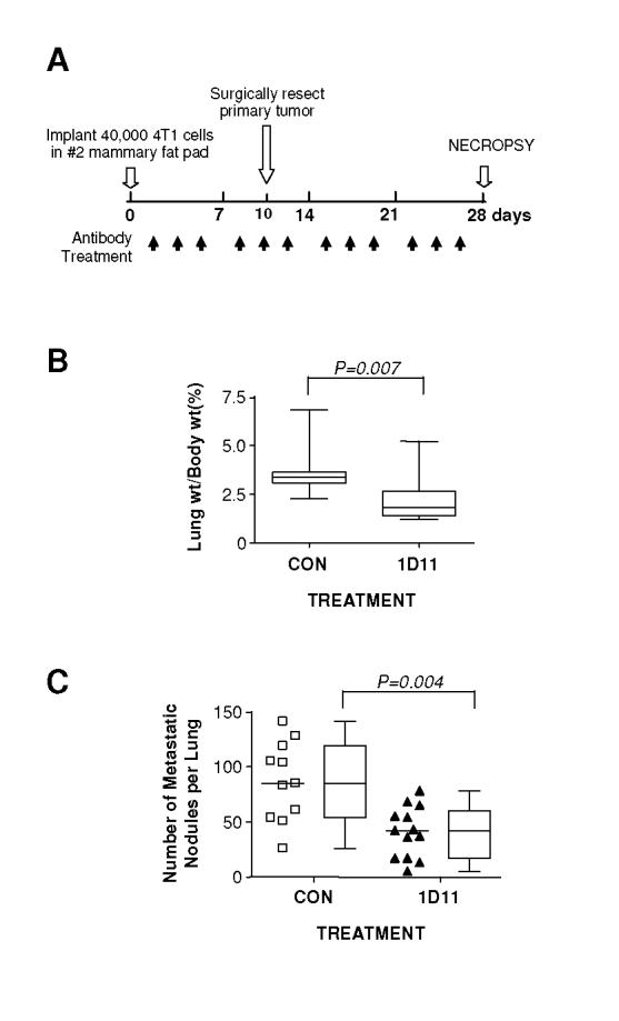
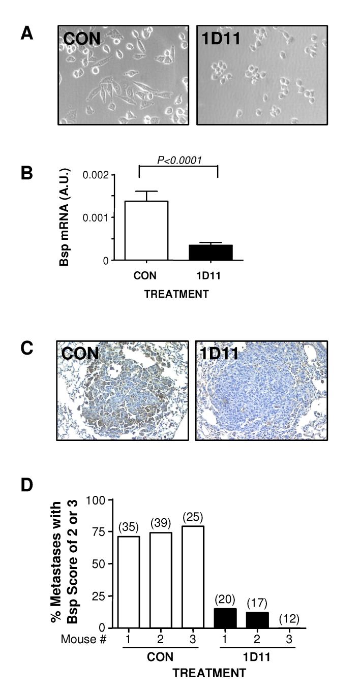
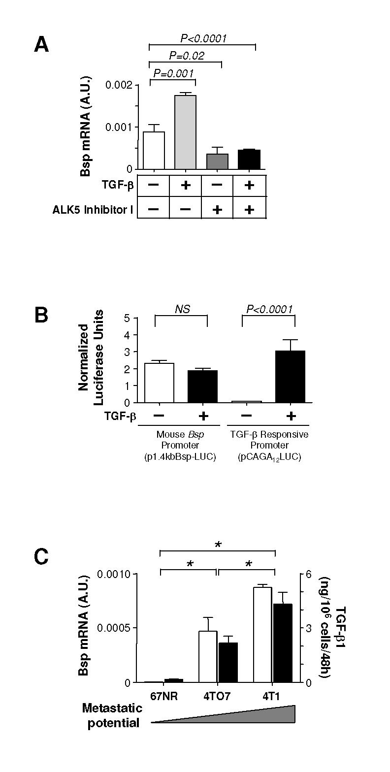
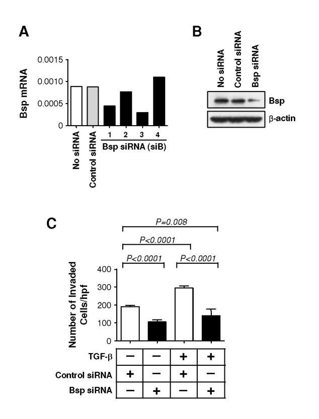
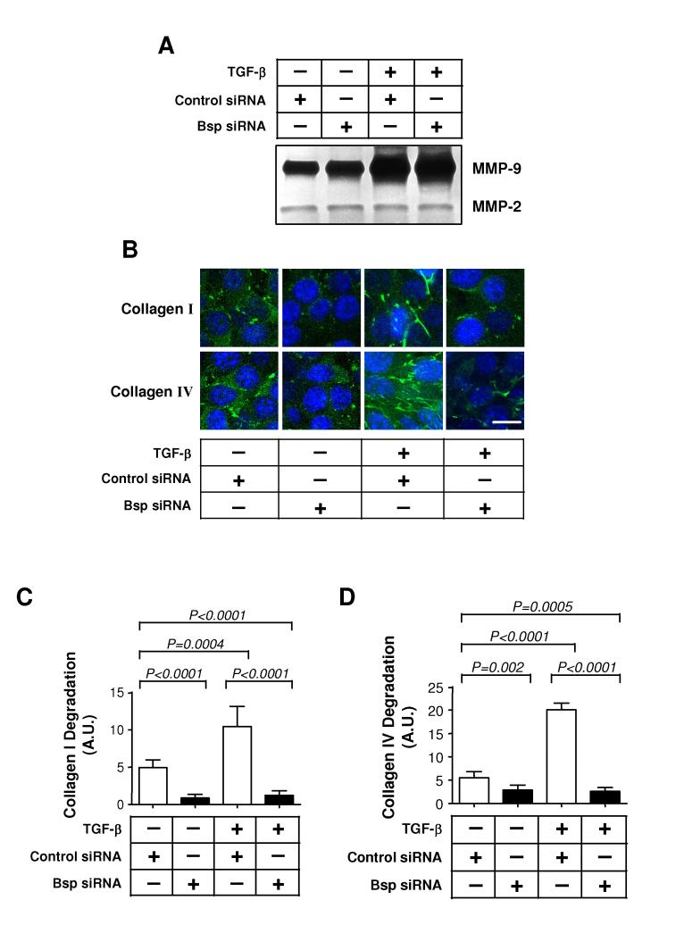
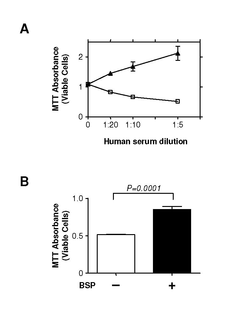
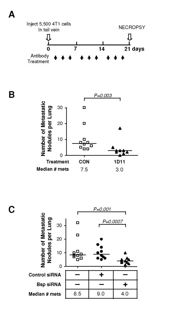
Transforming growth factor betas (TGF-beta) play a dual role in carcinogenesis, functioning as tumor suppressors early in the process, and then switching to act as prometastatic factors in late-stage disease. We have previously shown that high molecular weight TGF-beta antagonists can suppress metastasis without the predicted toxicities. To address the underlying mechanisms, we have used the 4T1 syngeneic mouse model of metastatic breast cancer. Treatment of mice with a monoclonal anti-TGF-beta antibody (1D11) significantly suppressed metastasis of 4T1 cells to the lungs. When metastatic 4T1 cells were recovered from lungs of 1D11-treated and control mice, the most differentially expressed gene was found to be bone sialoprotein (Bsp). Immunostaining confirmed the loss of Bsp protein in 1D11-treated lung metastases, and TGF-beta was shown to regulate and correlate with Bsp expression in vitro. Functionally, knockdown of Bsp in 4T1 cells reduced the ability of TGF-beta to induce local collagen degradation and invasion in vitro, and treatment with recombinant Bsp protected 4T1 cells from complement-mediated lysis. Finally, suppression of Bsp in 4T1 cells reduced metastasis in vivo. We conclude that Bsp is a plausible mediator of at least some of the tumor cell-targeted prometastatic activity of TGF-beta in this model and that Bsp expression in metastases can be successfully suppressed by systemic treatment with anti-TGF-beta antibodies.


Figures

Figure 1. 1D11 decreases 4T1 metastasis to the lung following orthotopic implantation.
A. Experimental schema. 4T1 cells (4 X 10⁴ cells) were inoculated into the left thoracic mammary fat pad of BALB/c mice. After the inoculation, either anti-TGF-β antibody 1D11 (5 mg/kg) or control 13C4 antibody, CON, (5 mg/kg) was administered three times per week i.p. to the mice. The primary tumors were removed on day 10 after the inoculation and mice were euthanized for necropsy on day 28. B. Relative lung weight at necropsy. C. Total number of grossly visible metastatic nodules in the lung. Boxes show median values with upper and lower quartiles, and whiskers indicate range (control group = 11 mice, 1D11 group = 13 mice).
Figure 2. Metastatic cells recovered from 1D11-treated lungs show persistent morphological changes in culture and elevated Bsp expression <i>in vitro</i> and <i>in vivo</i>.
Figure 2. Metastatic cells recovered from 1D11-treated lungs show persistent morphological changes in culture and elevated Bsp expression in vitro and in vivo.
4T1 cells were recovered from lungs harvested on day 28 as in Fig. 1 and cultured as described in Methods. A. Morphological appearance of the metastatic 4T1 cells cultured from the lungs of mice treated with anti-TGF-β (1D11) or control antibody (CON) after 7 days in culture. B. RTQ-PCR validation of differential expression of Bsp mRNA between metastatic cell cultures derived from treated and control mice. Values represent mean ± SD for 5 independent cell isolates/treatment group. Bsp mRNA expression was normalized to the 28S rRNA in each case. C. Immunohistochemical staining for Bsp in lung metastases from treated and control mice. D. Graphical representation of semi-quantitative scoring for Bsp expression in lung metastases. Individual metastases were scored for Bsp expression on a scale of 0-3 as detailed in methods. Data represent the % metastases with a score of ⩾ 2 for each of 3 mice/treatment group, for a total of 152 metastases evaluated. The number of metastases/mouse is indicated in parentheses above the bar.
Figure 3. TGF-β regulates and is correlated with Bsp expression in 4T1-related cell lines <i>in vitro</i>.
Figure 3. TGF-β regulates and is correlated with Bsp expression in 4T1-related cell lines in vitro.
A. Effect of TGF-β1 or a TGF-β type I receptor kinase inhibitor on the expression of Bsp mRNA by 4T1 cells in vitro. Cells were exposed to either TGF-β1 (5 ng/ml) or the ALK5 inhbitor I (10 μM) for 48 hours prior to analyzing Bsp expression by RTQ-PCR. B. Effect of TGF-β treatment on transcription from the mouse Bsp promoter was assessed by measuring luciferase activity in 4T1 cells transiently transfected with promoter-reporter constructs. P1.4kbBsp-LUC is the mouse Bsp promoter-luciferase construct and pCAGA12LUC is a synthetic TGF-β-responsive promoter-luciferase construct. Cells were treated with 5 ng/ml TGF-μ1 for 18 hours prior to assay. NS, no significance. C. Correlation between endogenous Bsp and TGF-β expression. Bsp mRNA expression ([box1]) was determined by RTQ-PCR, and secreted total (latent plus active) TGF-β1 protein levels ([square7]) were determined by ELISA assay in a series of 4T1-related cells of increasing metastatic potential. *P < 0.001. All values represent the mean ± SD for 3 determinations.
Figure 4. Bsp knockdown suppresses basal and TGF-β-induced invasion through Matrigel.
A. RTQ-PCR analysis of Bsp expression following transfection of 4T1 cells with four different siRNA sequences targeting Bsp. An siRNA to GFP was used as a negative control. B. Western blot analysis of Bsp protein levels in 4T1 cells 48 hours following transfection with the siB3 Bsp siRNA. C. Effect of Bsp knockdown on invasiveness of 4T1 cells. 4T1 cells transfected with siB3 were assessed for their ability to invade through Matrigel using a Transwell assay system, with and without added TGF-β1 (5 ng/ml). Results are the mean ± SD for 3 determinations.
Figure 5. Bsp knockdown does not affect induction of MMPs by TGF-β, but does reduce local activation of matrix degrading enzymes.
A. MMP expression assessed by conventional zymography. The effect of Bsp knockdown on basal and TGF-β-induced MMP expression was determined by zymography on gelatin-impregnated polyacrylamide gels of conditioned medium harvested from cells, cultured on type I collagen, following treatment with 5 ng/ml TGF-β1 or vehicle for 48 hours. B. Effect of Bsp knockdown on local matrix degradation as assessed by in-situ zymography of cells grown within agarose impregnated with type I or type IV DQ collagen. 4T1 cells transfected with Bsp siRNA or control siRNA were treated with TGF-β1 for 48 hours and analyzed by confocal microscopy. Local matrix degradation is visualized by release of the green fluorophore. Cell nuclei are visualized by DAPI staining. Scale bar = 20 μm C and D. Quantitation of extracellular matrix degradation in the in-situ zymography assay. The intensity of green fluorescence, representing degraded collagen, was quantitated for 9 random high power fields and normalized to the number of cells within the field, determined by DAPI staining (Blue), as detailed in Methods. C, type I collagen matrix; D, type IV collagen matrix.
Figure 6. Bsp protects 4T1 cells from complement-mediated cell lysis.
A. 4T1 cells are susceptible to complement-mediated cell lysis. 4T1 cells (5 X 10⁶ cells/ml) were suspended in GVB-MgEGTA buffer and incubated with different concentrations of normal human serum, with ([triangleup]) or without ([box1]) prior heat treatment to inactivate the complement. After 2 hours, cell viability was evaluated by colorimetric MTT assay. B. Bsp protects against complement-mediated lysis. 4T1 cells were incubated with purified human BSP protein (10 μg/ml) for 10 minutes prior to incubation with normal human serum diluted 1:5 for the lysis assay as above. Results are the mean ± SD for 3 determinations.
Figure 7. Knockdown of Bsp suppresses metastasis of 4T1 cells to the lung.
A. Experimental schema. 4T1 cells (5.5 X 10³ cells) were inoculated into tail vein of BALB/c mice. Where relevant, either 1D11 (5 mg/kg) or control 13C4 antibody, CON, (5 mg/kg) was administered three times per week i.p., starting one day after cell innoculation. Mice were euthanized for necropsy on day 21. B. Effect of 1D11 on metastatic efficiency. Mice were injected with 4T1 cells and then treated with 1D11 or control antibody. Grossly visible lung metastases were quantitated at necropsy range (control group = 10 mice, 1D11 group = 9 mice). C. Effect of Bsp knockdown on metastatic efficiency. Mice were injected with parental 4T1 cells, or 4T1 cells that had been transfected with either Bsp siRNA or control siRNA. Metastases were assessed as in B. All experimental groups contained 10 mice. Median numbers of metastases for each group are indicated (n = 10 for each group).

Similar articles

Cited by

References

    1. Derynck R, Akhurst RJ, Balmain A. TGF-β signaling in tumor suppression and cancer progression. Nat Genet. 2001;29:117–29. - PubMed
    1. Gold LI. The role for transforming growth factor-β (TGF-β) in human cancer. Crit Rev Oncog. 1999;10:303–60. - PubMed
    1. Wakefield LM, Roberts AB. TGF-β signaling: positive and negative effects on tumorigenesis. Curr Opin Genet Dev. 2002;12:22–9. - PubMed
    1. Yang YA, Dukhanina O, Tang B, et al. Lifetime exposure to a soluble TGF-β antagonist protects mice against metastasis without adverse side effects. J Clin Invest. 2002;109:1607–15. - PMC - PubMed
    1. Muraoka RS, Dumont N, Ritter CA, et al. Blockade of TGF-β inhibits mammary tumor cell viability, migration, and metastases. J Clin Invest. 2002;109:1551–9. - PMC - PubMed

Publication types

MeSH terms