. 2006 Jun;21(3):478-84.
doi: 10.3346/jkms.2006.21.3.478.

Disease-specific proteins from rheumatoid arthritis patients

Affiliations

Disease-specific proteins from rheumatoid arthritis patients

Choong Won Kim et al. J Korean Med Sci. 2006 Jun.

Abstract

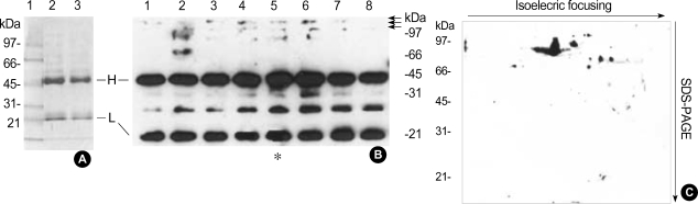
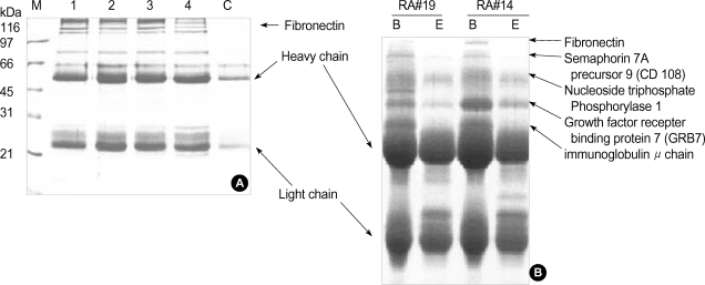
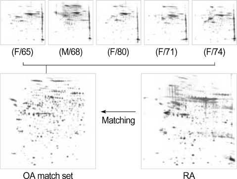
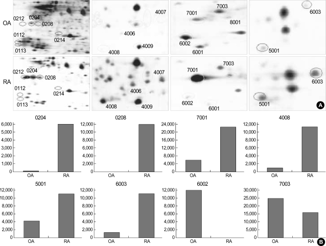
Rheumatoid arthritis (RA) is a chronic inflammatory disease that mainly destroys cartilages or bones at the joints. This inflammatory disorder is initiated by self-attack using own immune system, but the detail of pathological mechanism is unclear. Features of autoantigens leading to autoimmune disease are also under veil although several candidates including type II collagen have been suggested to play a role in pathogenesis. In this report, we tried to identify proteins responding to antibodies purified from RA patients and screen proteins up-regulated or down-regulated in RA using proteomic approach. Fibronectin, semaphorin 7A precursor, growth factor binding protein 7 (GRB7), and immunoglobulin mu chain were specifically associated with antibodies isolated from RA synovial fluids. In addition, some metabolic proteins such as adipocyte fatty acid binding protein, galectin-1 and apolipoprotein A1 precursor were overexpressed in RA synovium. Also, expression of peroxiredoxin 2 was up-regulated in RA. On the contrary, expression of vimentin was severely suppressed in RA synoviocytes. Such findings might give some insights into understanding of pathological mechanism in RA.


Figures

Fig. 1
Fig. 1
Purification of autoantibodies and blotting. (A) Antibody purification. Antibodies were purified from RA patients' synovial fluid using protein A affinity chromatography as described in "Materials and Methods". Two samples of antibody (each 10 µg) was analyzed by 12.5% SDS-PAGE. Lane 1 is protein size marker. H and L indicate heavy chain and light chain of immunoglobulins, respectively. (B) Blotting on a one-dimensional gel. Total cellular proteins (20 µg each lane) derived from RA synovium tissue (63/M) were separated on a 12.5% SDS-gel electrophoresis. They were subsequently transferred to the nitrocellulose membrane. Eight different primary antibodies isolated from RA patients (1:500 in 5% skim milk/TBST) were incubated with the membrane for 2 hr at room temperature using multi-channel blotting system (Bio-RAD). Secondary antibody, rabbit anti-human IgG HRP (1:2,000 in 5% skim milk/TBST), was incubated with membrane at room temperature for 1 hr. Proteins were visualized by a ECL developing system. (C) Blotting on a two-dimensional gel. Total proteins 150 µg from the RA synovium protein (63/M) was separated on a 7 cm and pH3-10 IPG strip, in the first dimension and 12.5% SDS-PAGE at the second dimension and subsequently transferred to the nitrocellulose membrane. Primary antibody from a RA patient (40/F) (1:100 in 5% skim milk/TBST) and subsequent HRP-conjugated rabbit anti-human IgGs (1:1,000 in 5% skim milk/TBST) were incubated with the membrane. Proteins were visualized by a ECL detecting system.
Fig. 2
Fig. 2
Isolation of autoantogens. (A) Affinity purification of autoantigens. Four different purified IgGs coupled to CNBr-activated Sepharose 4B were incubated with total synovial proteins for 1 hr at room temperature. After wash with binding buffer three times, one bead volume of 2X SDS sample buffer was added to protein-bound resins. After boiling for 5 min, the supernatant was separated on a 10% SDS-PAGE. C is a control, no cross-linked antibody. M indicates protein size marker. (B) Analysis of antigen-antibody complexes in synovial fluids. Synovial fluid (RA patients #14 and 19) was incubated with protein A resins. After washing protein A bead with 10 mM Tris-HCl (pH 7.5), beads were mixed with one bead volume of 2X SDS sample buffer and boiled for 7 min. The solubilized proteins were separated by a 10% SDS-PAGE after centrifugation at 13,000 rpm for 5 min and visualized by Coomassie blue staining. The specific proteins were identified by MALDI-TOF mass spectrometry as described (8-10). Lane B indicates antibody-antigen mix isolated and solubilized by SDS-sample buffer, and lane E is antibody sample eluted by low pH buffer as described in antibody purification.
Fig. 3
Fig. 3
An OA reference set for RA protein comparison. Total OA synovium proteins (40 µg) were separated on a two-dimensional gel: a linear IPG strip 17 cm and pH 4-7 at the first dimension and a 7.5-17.5% gradient SDS-PAGE at the second dimension. Protein spots were detected by silver staining. Five OA gels were matched together to create a reference set using the PDQuest program. This reference set was used for comparison with RA gels. Numbers in parentheses indicate age of patients.
Fig. 4
Fig. 4
Comparison of RA and OA proteins. Total RA or OA synovium proteins (40 µg) were separated on a two-dimensional gel: a linear IPG strip 17 cm and pH 4-7 at the first dimension and a 7.5-17.5% gradient SDS-PAGE at the second dimension. The RA gel was matched with the OA reference set (Fig. 3), and the relative values in (B) were obtained from the PDQuest analysis as described (8). The spot numbers were randomly given by the program.

References

    1. Ziff M. Rheumatoid arthritis-Its present and future. J Rheumatol. 1990;17:127–133. - PubMed
    1. Janossy G, Panayi G, Duke O, Bofill M, Poulter LW, Goldstein G. Rheumatoid arthritis: a disease of T-lymphocyte/macrophage immunoregulation. Lancet. 1981;2:839–842. - PubMed
    1. Cush JJ, Lipsky PE. Phenotypic analysis of synovial tissue and peripheral blood lymphocytes isolated from patients with rheumatoid arthritis. Arthritis Rheum. 1988;31:1230–1238. - PubMed
    1. Yamanishi Y, Firestein GS. Pathogenesis of rheumatoid arthritis: the role of synoviocytes. Rheum Dis Clin North Am. 2001;27:335–371. - PubMed
    1. Feldmann M, Brennan FM, Mainin RN. Rheumatoid arthritis. Cell. 1996;85:307–310. - PubMed

Publication types