

Langerhans cell sarcoma arising from Langerhans cell histiocytosis: a case report
- PMID: 16778410
- PMCID: PMC2729972
- DOI: 10.3346/jkms.2006.21.3.577
Langerhans cell sarcoma arising from Langerhans cell histiocytosis: a case report
Abstract
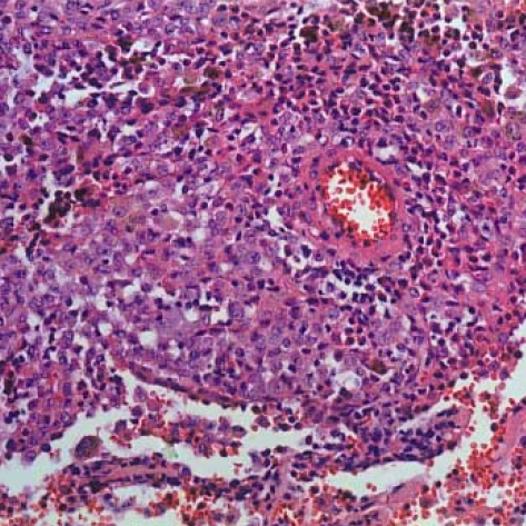
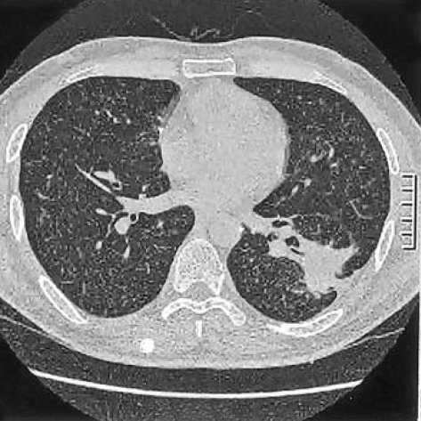
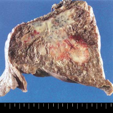
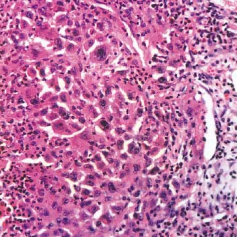
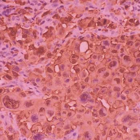
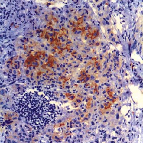
Langerhans cell sarcoma (LCS) is a neoplastic proliferation of Langerhans cells that have overtly malignant cytologic features. It is a very rare disease and theoretically, it can present de novo or progress from an antecedent Langerhans cell histiocytosis (LCH). However, to our knowledge, LCS arising from an antecedent LCH has not been reported on. We present here a case of LCS arising from a pulmonary LCH. A 34 yr-old man who was a smoker, had a fever and a chronic cough. Computed tomographic (CT) scan revealed multiple tiny nodules in both lungs. The thoracoscopic lung biopsy revealed LCH. The patient quit smoking, but he received no other specific treatment. One year later, the follow up chest CT scan showed a 4 cm-sized mass in the left lower lobe of the lung. A lobectomy was then performed. Microscopic examination of the mass revealed an infiltrative proliferation of large cells that had malignant cytologic features. Immunohistochemical stains showed a strong reactivity for S-100 and CD68, and a focal reactivity for CD1a. We think this is the first case of LCS arising from LCH.
Figures
References
-
- Jaffe ES, Harris NL, Stein H, Vardiman JW. WHO classification of tumours: Pathology and genetics of tumours of haematopoietic and lymphoid tissues. Lyon: IARCPress; 2001. p. 283.
-
- Ben-Ezra J, Bailey A, Azumi N, Delsol G, Stroup R, Sheibani K, Rappaport H. Malignant histiocytosis X. A distinct clinicopathologic entity. Cancer. 1991;68:1050–1060. - PubMed
-
- Imamura M, Sakamoto S, Hanazono H. Malignant histiocytosis: a case of generalized histiocytosis with infiltration of Langerhans' granule-containing histiocytes. Cancer. 1971;28:467–475. - PubMed
-
- Henderson DW, Sage RE. Malignant histiocytosis with eosinophilia. Cancer. 1973;32:1421–1428. - PubMed
-
- Wood C, Wood GS, Deneau DG, Oseroff A, Beckstead JH, Malin J. Malignant histiocytosis X. Report of a rapidly fatal case in an elderly man. Cancer. 1984;54:347–352. - PubMed
Publication types
MeSH terms
Substances
LinkOut - more resources
Full Text Sources
Medical
