. 2006 Jul 12;25(13):3033-44.
doi: 10.1038/sj.emboj.7601192. Epub 2006 Jun 15.

Adaptor functions of Cdc42, Ste50, and Sho1 in the yeast osmoregulatory HOG MAPK pathway

Affiliations

Adaptor functions of Cdc42, Ste50, and Sho1 in the yeast osmoregulatory HOG MAPK pathway

Kazuo Tatebayashi et al. EMBO J. .

Abstract

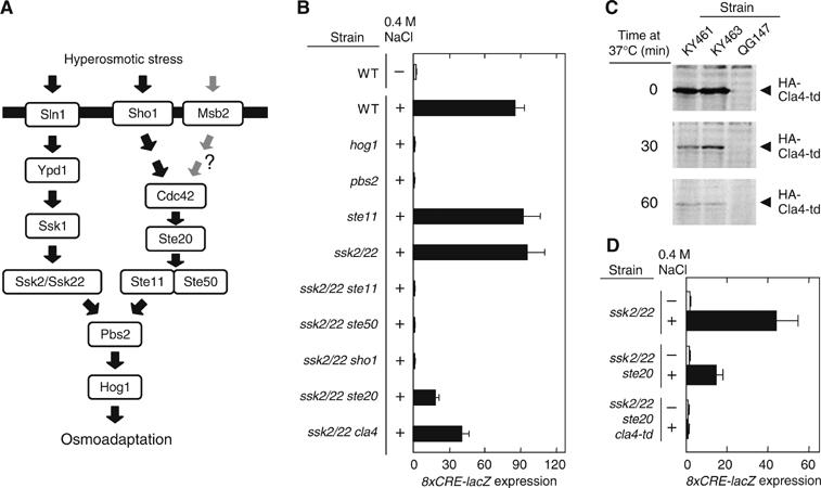
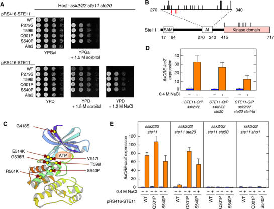
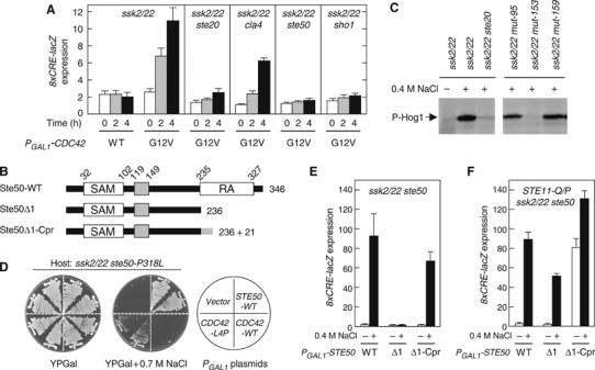
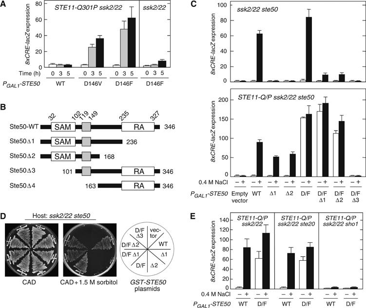
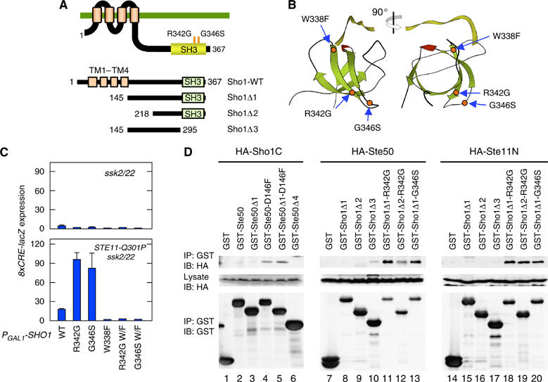
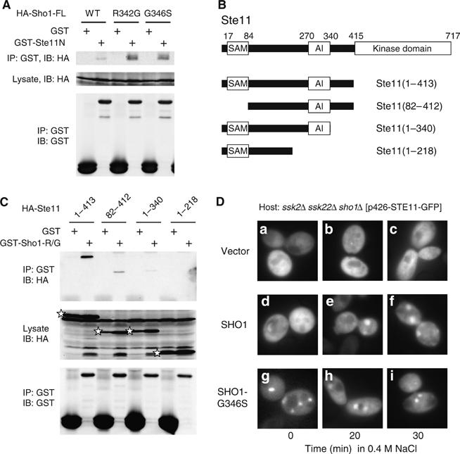
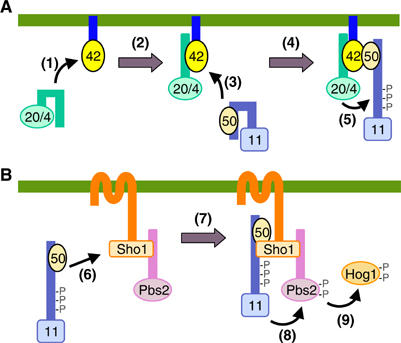
The yeast high osmolarity glycerol (HOG) signaling pathway can be activated by either of the two upstream pathways, termed the SHO1 and SLN1 branches. When stimulated by high osmolarity, the SHO1 branch activates an MAP kinase module composed of the Ste11 MAPKKK, the Pbs2 MAPKK, and the Hog1 MAPK. To investigate how osmostress activates this MAPK module, we isolated both gain-of-function and loss-of-function alleles in four key genes involved in the SHO1 branch, namely SHO1, CDC42, STE50, and STE11. These mutants were characterized using an HOG-dependent reporter gene, 8xCRE-lacZ. We found that Cdc42, in addition to binding and activating the PAK-like kinases Ste20 and Cla4, binds to the Ste11-Ste50 complex to bring activated Ste20/Cla4 to their substrate Ste11. Activated Ste11 and its HOG pathway-specific substrate, Pbs2, are brought together by Sho1; the Ste11-Ste50 complex binds to the cytoplasmic domain of Sho1, to which Pbs2 also binds. Thus, Cdc42, Ste50, and Sho1 act as adaptor proteins that control the flow of the osmostress signal from Ste20/Cla4 to Ste11, then to Pbs2.


Figures

Figure 1
Figure 1
Ste20 and Cla4 are redundant in the HOG pathway. (A) Schematic model of the yeast HOG signal pathway. The horizontal bar represents the plasma membrane. The role of Msb2 in HOG pathway is unclear (O'Rourke and Herskowitz, 2002; Cullen et al, 2004). (B) 8xCRE-lacZ expression accurately reflects the osmotic activation of the HOG pathway. Wild-type (TM141) and its derivatives with the indicated genotypes were transformed with the pRS414-8xCRE-lacZ reporter plasmid. β-Galactosidase activity was assayed before (−) or after (+) 30 min exposure to 0.4 M NaCl. Strains used are TM141, QG137, TM260, FP54, TM257, FP75, FP67, QG153, QG147, and QG148. (C) Degradation of Cla4-td at a nonpermissive temperature. KY461 (ssk2/22 ste20 cla4-td), KY463 (STE11-Q301P ssk2/22 ste20 cla4-td), and QG147 (ssk2/22 ste20) were grown exponentially at 25°C, transferred to 37°C, and at the indicated times, cell extracts were prepared. The HA-tagged Cla4-td protein was detected by immunoblotting using anti-HA antibody. (D) Ste20 and Cla4 are redundant in the SHO1 branch. Exponentially growing cells of the indicated genotypes carrying the pRS414-8xCRE-lacZ reporter plasmid were transferred from 25°C to 37°C 1 h before osmostress was applied. β-Galactosidase activity was assayed before (−) or after (+) 30 min exposure to 0.4 M NaCl. Strains used are TM257, QG147, and KY461.
Figure 2
Figure 2
Characterization of constitutively active Ste11 mutants. (A) Suppression of ste20Δ by constitutively active STE11 mutations. KY218 (ssk2 ssk22 ste11 ste20) carrying pRS416-STE11 or its derivatives was spotted on the indicated plates. Ala3 is a triple mutant of Ste20 phosphorylation sites, S281A S285A T286A. (B) Distribution of the constitutively active STE11 mutations. Below is the schematic diagram of Ste11 showing the SAM, autoinhibitory (AI) and kinase domains. AI domain is expanded above the diagram. Each black vertical bar represents a constitutively active STE11 mutation. Short bars indicate one allele at the position, while longer bars indicate two different alleles. Red bars below the horizontal line indicate phosphorylation sites. (C) The predicted three-dimensional locations of the constitutively active Ste11 mutations in the kinase domain. The crystallographic structure of the human PAK1 kinase domain was used as a model (PDB ID=1F3M). (D) Constitutively active Ste11 mutant bypasses both Ste20 and Cla4. Exponentially growing cells of the indicated genotypes carrying the pRS414-8xCRE-lacZ reporter plasmid were transferred from 25°C to 37°C 1 h before osmostress was applied. β-Galactosidase activity was assayed before (−) or after (+) 30 min exposure to 0.4 M NaCl. Strains used are KT018, KT031, and KY463. (E) Constitutively active Ste11 mutants require Ste50 and Sho1 for 8xCRE-lacZ induction. Mutant strains of the indicated genotypes carrying a 8xCRE-lacZ reporter plasmid and pRS416-STE11 or its indicated derivative were grown exponentially and incubated for 30 min with (+) or without (−) 0.4 M NaCl, before preparation of cell extracts for β-galactosidase assay. Strains used are FP75, KY213, KY444, and KY452.
Figure 3
Figure 3
Interaction between Cdc42 and Ste50. (A) Induction of the HOG reporter by the constitutively active Cdc42-G12V. Strains of the indicated genotypes were transformed with an 8xCRE-lacZ reporter plasmid and pYES2 (a multicopy plasmid with the GAL1 promoter) encoding either the wild-type Cdc42 (WT) or Cdc42-G12V. Expression of the Cdc42 protein was induced for 0, 2, or 4 h by 2% galactose, before preparation of cell extracts for β-galactosidase assay. Strains used are TM257, QG147, QG148, FP67, and QG153. (B) Schematic diagram of Ste50. Boxes indicate the SAM and RA domains and a central conserved region. Deletion (Δ1) and membrane-targeting (Δ1-Cpr) mutants are also shown. Cpr, C-terminal prenylation signal of Ras2. (C) The mut-153 (ste50-P318L) strain is defective in Hog1 phosphorylation. Exponentially growing cells were stimulated with 0.4 M NaCl for 5 min before cell extracts were prepared. Phosphorylated Hog1 was probed by immunoblotting using anti-phospho p38 antibody. Control cells on the left panel are TM252 and FP53. mut-95 and mut-159 are irrelevant control mutants isolated in the same screening. (D) Suppression of the osmosensitivity of ste50-P318L mutant cells by CDC42-L4P. FP67 (ssk2 ssk22 ste50) carrying pRS416-ste50-P318L was transformed with YCpIF16 (a single-copy vector with the GAL1 promoter) encoding the indicated gene, and streaked on YPGal plates with or without 0.7 M NaCl. (E, F) A membrane-targeting signal functionally substitutes for the Ste50 RA domain. Host strains of the indicated genotypes (FP67 and KT049) carrying pRS416-8xCRE-lacZ were transformed with YCpIF16-STE50 or its mutant derivatives. Cells were grown exponentially in CARaf, induced for Ste50 expression for 1.5 h by adding galactose to 2%, and incubated for an additional 30 min with (+) or without (−) 0.4 M NaCl in the media.
Figure 4
Figure 4
Constitutively active Ste50 mutant. (A) Constitutively active Ste50 mutants. KT018 (STE11-Q301P ssk2 ssk22) and TM257 (ssk2 ssk22) carrying the pRS416-8xCRE-lacZ reporter plasmid were transformed with YCpIF16-STE50 or its derivative as indicated. Ste50 expression was induced by 2% galactose for 0, 3, and 5 h before cell extracts were prepared for β-galactosidase assay. (B) Schematic diagrams of the Ste50 deletion constructs used in this work. (C) Induction of the 8xCRE-lacZ reporter by the constitutively active Ste50-D146F mutant. FP67 (ssk2 ssk22 ste50) and KT049 (STE11-Q301P ssk2 ssk22 ste50) carrying the pRS416-8xCRE-lacZ reporter plasmid were transformed with YCpIF16-STE50 or one of its derivatives containing the indicated mutations. Cells were grown exponentially in CARaf, induced for Ste50 expression for 1.5 h by 2% galactose, and incubated for additional 30 min with (+) or without (−) 0.4 M NaCl in the media, before cell extracts were prepared for β-galactosidase assay. D/F, D146F. (D) Intragenic suppression of the Ste50 RA domain deletion mutants by D146F. FP67 (ssk2 ssk22 ste50) was transformed with YCpIF16 (a single-copy vector with the GAL1 promoter) encoding the indicated gene, and streaked on CAD plates with or without 1.5 M sorbitol. (E) Constitutively active Ste50-D146F requires Sho1 for 8xCRE-lacZ reporter expression. Cells of the indicated genotypes (KT018, KT031, and KT028) carrying pRS416-8xCRE-lacZ were transformed with either YCpIF16-Ste50 (WT) or YCpIF16-Ste50-D146F (D/F). Cell extracts for β-galactosidase assay were prepared as in (C).
Figure 5
Figure 5
Constitutively active Sho1 mutants. (A) Schematic diagrams of the Sho1 mutants used in this work. TM, transmembrane segment. (B) The predicted three-dimensional locations of the constitutively active Sho1 mutations R342G and G346S in the SH3 domain. The location of W338F that abrogates Pbs2 binding is also shown. The crystallographic structure of the human Fyn SH3 domain (green) complexed with a ten-residue proline-rich peptide (yellow) was used as a homologous model (PDB ID=1FYN). (C) Constitutively active Sho1 mutants induce 8xCRE-lacZ reporter expression in the presence of Ste11-Q301P. TM257 (ssk2 ssk22) and KT018 (STE11-Q301P ssk2 ssk22) carrying the pRS414-8xCRE-lacZ reporter plasmid were transformed with pRS416-GAL1-SHO1 or one of its derivatives containing the indicated mutations. The Sho1 proteins were induced by 2% galactose for 2 h before cell extracts were prepared for β-galactosidase assay. W/F, W338F. (D) Binding of Sho1 to Ste50 and Ste11. KT045 (sho1 ste11 ste50 pbs2) was co-transformed with a YCpIF16-based plasmid (GAL1 promoter) for expression of HA-tagged protein and a pTEG1-based plasmid (TEF promoter) for expression of GST-fusion protein. HA-Sho1C includes residues 145–367, HA-Ste50 contains the entire Ste50 coding region (1–346), and HA-Ste11N contains residues 1–413. Cells were grown in CARaf medium, and expression of the fusion proteins was induced for 4 h by 2% galactose. GST-fusion proteins were captured by glutathione-Sepharose beads, and co-precipitated HA-tagged protein was detected by immunoblotting.
Figure 6
Figure 6
Interaction between Sho1 and Ste11. (A) Binding of the full-length Sho1 to Ste11. TM141 (wild-type) cells were co-transformed with a YCpIF16-based plasmid that encodes HA-tagged full-length Sho1 protein with the indicated mutation, and pTEG1-STE11N, which encodes GST-Ste11N. See the Figure 5D legend for other details. (B) Schematic diagram of the Ste11 deletion constructs used for the co-immunoprecipitation assays in (C). Boxes indicate the SAM, AI, and kinase catalytic domains. (C) Binding of Ste11 N-terminal fragments to Sho1C-R342G. KT045 (sho1 ste11 ste50 pbs2) was co-transformed with a YCpIF16-based plasmid (GAL1 promoter) for expression of HA-tagged Ste11 N-terminal fragments as indicated, and a pTEG1-based plasmid (TEF promoter) for expression of GST-Sho1C-R342G (R/G) or GST alone. Cells were grown in CARaf medium, and expression of the fusion proteins was induced for 4 h by 2% galactose. GST-fusion proteins were captured by glutathione-Sepharose beads, and co-precipitated HA-tagged protein was detected by immunoblotting. The positions of HA-Ste11 proteins in the total lysate are indicated by stars: other bands are background. (D) QG153 (ssk2 ssk22 sho1) carrying pRS426-Ste11-GFP (in which Ste11-GFP is expressed under the control of the STE11 promoter) was co-transformed with a pRS414-based plasmid encoding either Sho1-WT or Sho1-G346S under the control of the GAL1 promoter. Cells were grown in CARaf to log phase, galactose was added to a final concentration of 2%, grown for 3 h, and stimulated with 0.4 M NaCl for indicated time. Cells were kept at the room temperature (23°C) after the NaCl addition.
Figure 7
Figure 7
Schematic model of the sequential docking interactions in the SHO1 pathway. (A) Activation of Ste11 by Ste20 is mediated by indirect docking via Ste50 and Cdc42. (B) Activation of Pbs2 by Ste11 is mediated by indirect docking via Ste50 and Sho1. The green horizontal bars represent the plasma membrane. 11, Ste11; 20/4, Ste20/Cla4; 42, Cdc42; 50, Ste50. The parenthesized numbers in the figure correspond to the step numbers in the text.

References

    1. Ash J, Wu C, Larocque R, Jamal M, Stevens W, Osborne M, Thomas DY, Whiteway M (2003) Genetic analysis of the interface between Cdc42p and the CRIB domain of Ste20p in Saccharomyces cerevisiae. Genetics 163: 9–20 - PMC - PubMed
    1. Bhattacharya S, Chen L, Broach JR, Powers S (1995) Ras membrane targeting is essential for glucose signaling but not for viability in yeast. Proc Natl Acad Sci USA 92: 2984–2988 - PMC - PubMed
    1. Brewster JL, de Valoir T, Dwyer ND, Winter E, Gustin MC (1993) An osmosensing signal transduction pathway in yeast. Science 259: 1760–1763 - PubMed
    1. Cullen PJ, Sabbagh W Jr, Graham E, Irick MM, van Olden EK, Neal C, Delrow J, Bardwell L, Sprague GF Jr (2004) A signaling mucin at the head of the Cdc42- and MAPK-dependent filamentous growth pathway in yeast. Genes Dev 18: 1695–1708 - PMC - PubMed
    1. Cvrckova F, De Virgilio C, Manser E, Pringle JR, Nasmyth K (1995) Ste20-like protein kinases are required for normal localization of cell growth and for cytokinesis in budding yeast. Genes Dev 9: 1817–1830 - PubMed

Publication types

MeSH terms

Substances