. 2006 Jul 12;25(13):3214-22.
doi: 10.1038/sj.emboj.7601189. Epub 2006 Jun 15.

Toxoplasma gondii scavenges host-derived lipoic acid despite its de novo synthesis in the apicoplast

Affiliations

Toxoplasma gondii scavenges host-derived lipoic acid despite its de novo synthesis in the apicoplast

Michael J Crawford et al. EMBO J. .

Abstract

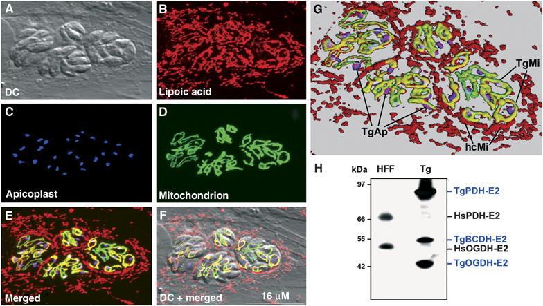
In contrast to other eukaryotes, which manufacture lipoic acid, an essential cofactor for several vital dehydrogenase complexes, within the mitochondrion, we show that the plastid (apicoplast) of the obligate intracellular protozoan parasite Toxoplasma gondii is the only site of de novo lipoate synthesis. However, antibodies specific for protein-attached lipoate reveal the presence of lipoylated proteins in both, the apicoplast and the mitochondrion of T. gondii. Cultivation of T. gondii-infected cells in lipoate-deficient medium results in substantially reduced lipoylation of mitochondrial (but not apicoplast) proteins. Addition of exogenous lipoate to the medium can rescue this effect, showing that the parasite scavenges this cofactor from the host. Exposure of T. gondii to lipoate analogues in lipoate-deficient medium leads to growth inhibition, suggesting that T. gondii might be auxotrophic for this cofactor. Phylogenetic analyses reveal the secondary loss of the mitochondrial lipoate synthase gene after the acquisition of the plastid. Our studies thus reveal an unexpected metabolic deficiency in T. gondii and raise the question whether the close interaction of host mitochondria with the parasitophorous vacuole is connected to lipoate supply by the host.


Figures

Figure 1
Figure 1
Detection of lipoylated proteins in the apicoplast and mitochondrion of T. gondii and host cell mitochondria by confocal immunofluorescence microscopy and immunoblot analysis. Infected HFF cells (A) were stained with polyclonal anti-LA antibody and Cy3-labeled secondary antibody (B). Apicoplast-resident HA-FNR was visualized using anti-HA and secondary Cy5-labeled antibodies (C). The mitochondrion could be detected by GFP fluorescence (see Materials and methods) (D). In (E) and (F), all three fluorescent channels are merged. Pink color indicates colocalization of lipoylated proteins with the apicoplast, yellow with the mitochondrion of the parasites. (G) Represents a partial blow-up of (E), which was also volume-rendered using VolumeJ software. The background color was changed to gray for better visualization of organelles. Some host cell mitochondria surrounding the parasitophorous vacuolar membrane are indicated (hcMi). Parasite mitochondria (TgMi) and apicoplasts (TgAp) are also outlined. Note the close association between apicoplast and mitochondrion. (H) Immunoblot analysis of uninfected host cells (HFF) and purified T. gondii tachyzoites (Tg) with polyclonal anti-LA antibody. The positions and names of host (Hs) and parasite (Tg) lipoylated proteins reacting with this antibody are indicated (see text for details).
Figure 2
Figure 2
Reduction of apicoplast, but not mitochondrial, lipoylation upon triclosan treatment. (A) Untreated tachyzoites 16 h postinfection, (B) triclosan-treated parasites after 36 h in the presence of 0.3 μg/ml triclosan. The merged images are composites of mitochondria (green), polyclonal anti-LA (red), and apicoplast (blue). The whole color plate with individual images can be found in Supplementary Figure S-8. (C, D) Enlarged images of the dashed areas in (A and B), respectively. In three exemplary regions, circles indicate the presence of colocalization of lipoylated proteins within the apicoplast (C, pink color) or its absence due to triclosan treatment (D, only blue color). (E) Immunoblot analysis of lipoylated proteins from purified T. gondii from untreated or triclosan-treated samples (0.3 μg/ml for 48 h) using polyclonal anti-LA antibody.
Figure 3
Figure 3
Complementation of the triclosan-mediated lipoylation defect by recombinant overexpression of T. gondii enoyl-ACP reductase (TgENR). A stable T gondii clone recombinantly expressing the predicted triclosan target (apicoplast TgENR) with a C-terminal HA tag (see Materials and methods) was created. Immunoblot analysis with anti-LA antibody probed against lysates of purified T. gondii (ptubTgENR-HA) and wild-type parasites (RH) treated with the indicated triclosan concentrations for 48 h reveals the specific effect of TgENR overexpresssion on TgPDH-E2 lipoylation in triclosan-treated cultures.
Figure 4
Figure 4
Lipoylation of mitochondrial T. gondii proteins in LA-deficient medium. HFF infected with RHβ1 parasites were cultivated in LAM5 in the absence or presence of 1 μM exogenous LA for 48 h and then analyzed by SDS–PAGE and immunoblotting with polyclonal anti-LA antibodies (lanes 1–2). The same experiment was also performed in the presence of 0.5 μg/ml of triclosan (lanes 3–4). Mitochondrial parasite proteins TgBCDH-E2 and TgOGDH-E2 showed greatly reduced lipoylation signals in the absence of LA (lane 2). In addition, lipoylation of TgPDH-E2 was reduced in the absence of fatty acid synthesis (lane 3). Parasite-encoded β-galactosidase (TgβGal) and the 70 kDa subunit of human mitochondrial succinate-ubiquinol oxidoreductase (complex II; HsSDHA), respectively, served as loading control for each cell type.
Figure 5
Figure 5
Inhibitory effects of LA analogs on T. gondii growth and protein lipoylation. RHβ1 were cultured in LAM5 in the presence of the indicated concentrations of fatty acid analogs in the absence (striped bars) or presence of 1 μM LA (black bars) in 48-well plates for 72 h and then assayed for β-galactosidase activity as a means for parasite growth as described previously (Seeber and Boothroyd, 1996). Fatty acid abbreviations are given in the text. OD values of the untreated control were then set as 100% growth and the other values calculated accordingly. Note the consistent beneficial effect on growth by LA supplementation in untreated cells.
Figure 6
Figure 6
Maximum likelihood phylogenetic analysis of LipA protein sequences. Selected sequences representing major taxa whose genome sequences have been determined entirely were analyzed as described in Materials and methods. All eukaryotic sequences were analyzed for the presence of N-terminal organellar targeting sequences using various bioinformatic tools (see Supplementary Table S-I). Their known or predicted organellar location correlates very well with their phylogenetic clustering (Supplementary Table S-I). Numbers at the basis of the nodes are bootstrap values of 100 replicates. The different clades are outlined (cyanobacteria within the eubacterial clade are striped), the α-proteobacterium Rickettsia prowazekii is highlighted, and the two apicomplexan LipA's are boxed. Organism identification codes are according to the UniProt group.

References

    1. Ali ST, Moir AJ, Ashton PR, Engel PC, Guest JR (1990) Octanoylation of the lipoyl domains of the pyruvate dehydrogenase complex in a lipoyl-deficient strain of Escherichia coli. Mol Microbiol 4: 943–950 - PubMed
    1. Andersson SG, Zomorodipour A, Andersson JO, Sicheritz-Ponten T, Alsmark UC, Podowski RM, Naslund AK, Eriksson AS, Winkler HH, Kurland CG (1998) The genome sequence of Rickettsia prowazekii and the origin of mitochondria. Nature 396: 133–140 - PubMed
    1. Augustin S, Nolden M, Müller S, Hardt O, Arnold I, Langer T (2005) Characterization of peptides released from mitochondria: evidence for constant proteolysis and peptide efflux. J Biol Chem 280: 2691–2699 - PubMed
    1. Biewenga G, Haenen G, Bast A (1997) An overview of lipoate chemistry. In Lipoic Acid in Health and Disease, Fuchs J, Packer L, Zimmer G (eds) Vol. 6, pp 1–32. New York: Marcel Dekker
    1. Coppens I, Joiner KA (2001) Parasite–host cell interactions in toxoplasmosis: new avenues for intervention? Expert Rev Mol Med 2001: 1–20 - PubMed

Publication types