. 2006 Jul;116(7):2044-55.
doi: 10.1172/JCI27727. Epub 2006 Jun 15.

The IL-21 receptor augments Th2 effector function and alternative macrophage activation

Affiliations

The IL-21 receptor augments Th2 effector function and alternative macrophage activation

John Pesce et al. J Clin Invest. 2006 Jul.

Abstract

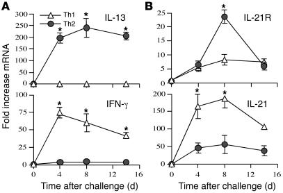
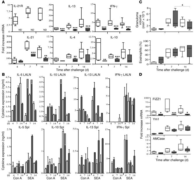
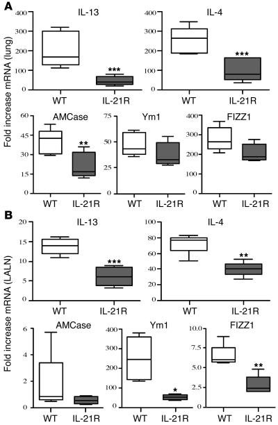
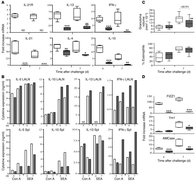
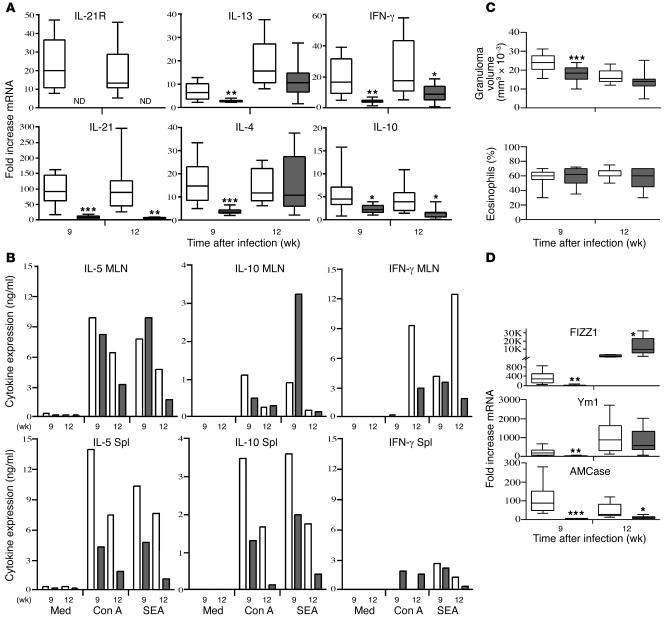
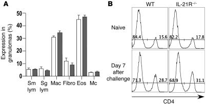
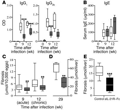
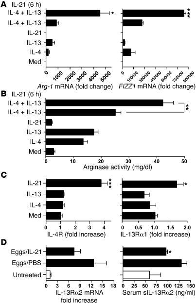
The IL-21 receptor (IL-21R) shows significant homology with the IL-4R, and CD4+ Th2 cells are an important source of IL-21. Here we examined whether the IL-21R regulates the development of Th2 responses in vivo. To do this, we infected IL-21R-/- mice with the Th2-inducing pathogens Schistosoma mansoni and Nippostrongylus brasiliensis and examined the influence of IL-21R deficiency on the development of Th2-dependent pathology. We showed that granulomatous inflammation and liver fibrosis were significantly reduced in S. mansoni-infected IL-21R-/- mice and in IL-21R+/+ mice treated with soluble IL-21R-Fc (sIL-21R-Fc). The impaired granulomatous response was also associated with a marked reduction in Th2 cytokine expression and function, as evidenced by the attenuated IL-4, IL-13, AMCase, Ym1, and FIZZ1 (also referred to as RELMalpha) responses in the tissues. A similarly impaired Th2 response was observed following N. brasiliensis infection. In vitro, IL-21 significantly augmented IL-4Ralpha and IL-13Ralpha1 expression in macrophages, resulting in increased FIZZ1 mRNA and arginase-1 activity following stimulation with IL-4 and IL-13. As such, these data identify the IL-21R as an important amplifier of alternative macrophage activation. Collectively, these results illustrate an essential function for the IL-21R in the development of pathogen-induced Th2 responses, which may have relevance in therapies for both inflammatory and chronic fibrotic diseases.


Figures

Figure 1
Figure 1. IL-21 and IL-21R expression during highly polarized Th1 and Th2 immune responses.
IL-10–/–IL-4–/– (Th1) and IL-10–/–IL-12–/– (Th2) mice (n = 5 per group) were sensitized i.p. with freshly isolated eggs of S. mansoni and challenged i.v. 14 days later. On days 4, 8, and 14 after challenge, mice were sacrificed, and lung RNA specimens were prepared individually for real-time RT-PCR analysis of IL-13 and IFN-γ (A) as well as IL-21 and IL-21R (B). Gene expression (mean ± SEM) is expressed as the fold increase over unchallenged WT controls after normalization to HPRT. Similar results were obtained in several repeat experiments. *P < 0.05.
Figure 2
Figure 2. Th2 cytokine production is reduced in the lungs of schistosome egg–challenged IL-21R–/– mice.
Groups of naive WT (white bars) and IL-21R–/– mice (gray bars) were i.v. challenged with live S. mansoni eggs and sacrificed on days 4, 7, and 14 after challenge. (A) RNA was prepared from lung tissues and analyzed individually (n = 5 per group/time point) by real-time RT-PCR. The top and bottom of the boxes indicate the seventy-fifth and twenty-fifth percentiles, respectively; the line within the box indicates the fiftieth percentile; and the top and bottom whiskers indicate the ninetieth and tenth percentiles, respectively, of the tested samples. ND, not detected. *P < 0.05, **P < 0.01, ***P < 0.001 versus WT. (B) Spleens (Spl) and lung-associated lymph nodes (LALN) were each pooled (2 separate groups, 3–4 mice per group), and single-cell suspensions were assayed for IL-5, IL-10, IL-13, and IFN-γ after a 72-hour incubation in the presence of Con A (1 mg/ml) or SEA (20 mg/ml). Results are mean ± SEM. Cytokines were below the level of detection in unstimulated cultures. (C) Granuloma size (volume, mm3 × 10–3) and the percentage of eosinophils in granulomas were quantified microscopically. (D) Real-time PCR analysis of Th2-regulated inflammatory genes in granulomatous lung tissue. All data are representative of at least 2 separate experiments.
Figure 3
Figure 3. The Th2 response is impaired in N. brasiliensis –infected IL-21R–/– mice.
Lungs (A) and lung-associated lymph nodes (B) were removed on day 7 from individual N. brasiliensis–infected and untreated C57BL/6 or IL-21R–/– mice (n = 5 per treatment group). RNA was isolated and cDNA was generated as described in the legend to Figure 2. mRNA was analyzed individually for IL-13, IL-4, AMCase, Ym1, and FIZZ1 by real-time quantitative PCR. Fold changes are based on comparisons of infected mice to naive animals. The experiment was repeated with similar results. *P < 0.05, **P < 0.01, ***P < 0.001 versus WT.
Figure 4
Figure 4. Th2 cytokine–driven inflammation is reduced in IL-21R–/– mice.
WT (white bars) and IL-21R–/– mice (gray bars) were sensitized i.p. with eggs, challenged i.v. 2 weeks later with live S. mansoni eggs, and then sacrificed on days 4 and 7 after challenge. (A) RNA was prepared from lung tissues and analyzed individually (n = 5 per group/time point) by real-time RT-PCR as described above in the legend to Figure 2. (B) Spleens and lung-associated lymph nodes were assayed for IL-5, IL-10, IL-13, and IFN-γ following antigen (SEA) or mitogen stimulation (Con A). (C) Granuloma size (mm3 × 10–3) and the percentage of eosinophils in granulomas were quantified microscopically in WT (n = 10 [day 4]; 15 [day 7]) and IL-21R–/– mice (n = 11 [day 4]; 16 [day 7]). (D) Real-time PCR analysis of Th2 inflammatory genes in granulomatous lung tissue (n = 5 per group/time point). Data shown are the combined results of 3 separate experiments. *P < 0.05, **P < 0.01, ***P < 0.001 versus WT.
Figure 5
Figure 5. Chronic liver disease following percutaneousS. mansoni infection is reduced in the absence of IL-21R.
WT (white bars) and IL-21R–/– mice (gray bars) were infected with 25–30 S. mansoni cercariae. All animals were sacrificed 9 (acute) or 12 (chronic) weeks after infection. (A) RNA was isolated from liver tissues and analyzed individually (n = 8–10 per group/time point) by real-time RT-PCR as described in the legend to Figure 2. (B) Spleens and mesenteric lymph nodes (MLN) were pooled in groups of 2–4 mice, and single-cell suspensions were assayed for IL-5, IL-10, and IFN-γ. The data shown are the averages of 3 separate pooled groups. Med, medium alone. (C) Granuloma size (mm3 × 10–3) and the percentage of eosinophils in granulomas were evaluated microscopically in WT (n = 30 [week 9]; 17 [week 12]) and IL-21R–/– mice (n = 27 [week 9]; 19 [week 12]). (D) Real-time PCR analysis of Th2 inflammatory genes in granulomatous liver tissue (n = 8–10 per group/time point). Data shown are the combined results of 3 separate experiments conducted on week 9 and 2 performed on week 12. *P < 0.05, **P < 0.01, ***P < 0.001 versus WT.
Figure 6
Figure 6. The cellular composition of granulomas is unchanged by IL-21R deficiency.
(A) The cellular composition of individual granulomas was evaluated in the livers of 9 week infected WT (white bars; n = 10) and IL-21R–/– (gray bars; n = 9) mice. The mean ± SEM of small lymphocytes (Sm lym; including the CD4/CD8 T cells), large lymphocytes (Lg lym; B cells and plasma cells), macrophages (Mac), fibroblasts (Fibro), eosinophils (Eos), and mast cells (Mc) are shown. (B) Lymphocytes were isolated from the perfused lungs of naive WT and IL-21R–/– mice and on day 7 following i.v. challenge with 5,000 S. mansoni eggs. The numbers in the histograms indicate the percent of CD4 and CD4+ T cells among total lung lymphocytes.
Figure 7
Figure 7. IL-21R deficiency significantly slows the progression of Th2 cytokine–dependent fibrosis.
WT (white bars) and IL-21R–/– mice (gray bars) were infected with S. mansoni cercariae. Animals were sacrificed 9 (acute) and 12 weeks (chronic; AC) or 29 weeks (late chronic; D) after infection. (A) Mice were bled at the time of sacrifice, and SEA isotype–specific Ab titers were determined by ELISA. (B) Total serum IgE values. (CE) Fibrosis was assessed by the amount of hydroxyproline (μmol) detected in the liver per 10,000 eggs (C) or per the total liver (D and E). In E, infected WT C57BL/6 mice were treated with either an IgG2a control Ab or with sIL-21R–Fc for 5 weeks. *P < 0.05, **P < 0.01, ***P < 0.001 versus WT.
Figure 8
Figure 8. IL-21 signaling promotes alternative macrophage activation by modulating IL-13R expression.
Bone marrow–derived macrophages were treated with IL-21 (20 ng/ml), either alone or in combination with IL-4 (20 ng/ml) and IL-13 (20 ng/ml) overnight. In some experiments, macrophages were pretreated with IL-21 for 6 hours prior to IL-4 and IL-13 administration. Cells were lysed 20 hours later, and RNA was analyzed individually by real-time RT-PCR. (A) The ability of IL-21 to promote alternative macrophage activation was assessed by measuring Arg-1 and FIZZ1 mRNA expression. (B) Arginase activity was quantified in cell lysates by measuring the conversion of l-arginine to Urea (mg/dl ± SEM; triplicate measurements). (C) Expression of IL-4Rα and IL-13Rα1 mRNA was evaluated by real-time PCR. IL-13Rα2 mRNA was nearly undetectable in all conditions (not shown). The data shown in A, B, and C are representative of 3 separate experiments. (D) Naive C57BL/6 mice were challenged i.v. with 5,000 live S. mansoni eggs and treated with PBS or recombinant IL-21 (2 μg/dose) every other day from day 1 through day 6. Animals (n = 5 per group) were sacrificed on day 7, and lung IL-13Rα2 mRNA levels were assayed by real-time PCR and expressed as fold increase over untreated controls. Mice were also bled at the time of sacrifice, and the amount of sIL-13Rα2 in individual serum samples was assayed by ELISA. *P < 0.05; **P < 0.01; ***P < 0.001. Similar results were obtained in a separate study.

References

    1. Parrish-Novak J., et al. Interleukin 21 and its receptor are involved in NK cell expansion and regulation of lymphocyte function. Nature. 2000;408:57–63. - PubMed
    1. Ozaki K., Kikly K., Michalovich D., Young P.R., Leonard W.J. Cloning of a type I cytokine receptor most related to the IL-2 receptor beta chain. Proc. Natl. Acad. Sci. U. S. A. 2000;97:11439–11444. - PMC - PubMed
    1. Sivakumar P.V., Foster D.C., Clegg C.H. Interleukin-21 is a T-helper cytokine that regulates humoral immunity and cell-mediated anti-tumour responses. Immunology. 2004;112:177–182. - PMC - PubMed
    1. Habib T., Nelson A., Kaushansky K. IL-21: a novel IL-2-family lymphokine that modulates B, T, and natural killer cell responses. J. Allergy Clin. Immunol. 2003;112:1033–1045. - PubMed
    1. Vosshenrich C.A., Di Santo J.P. Cytokines: IL-21 joins the gamma(c)-dependent network? Curr. Biol. 2001;11:R175–R177. - PubMed

Publication types

MeSH terms